SOP: Identification of Listeria monocytogenes using the Adiafood Rapid Pathogen Detection System, a Real-Time PCR Technique
GIFSQ — Global Institute for Food Safety & Quality
Principle
Following an initial 24 hour pre-enrichment that is common with the cultural method for the isolation of Listeria monocytogenes (MFHPB-30), an aliquot of the broth is transferred onto a Palcam agar slant for an additional 18 to 24 hours. The cell suspension obtained by washing the Palcam agar slant with peptone water is subjected to a real-time PCR procedure in which the target pathogen DNA is amplified and detected using specific primers and molecular beacons. Molecular beacons are unique sequence probes that allow for the identification of the pathogen with a high level of specificity. Once bound to their target, the molecular beacons emit a fluorescent signal that is proportional to the amount of amplified pathogenic DNA. In the absence of target bacteria in food samples, no fluorescent signal is detected, since the oligonucleotide primers used within this system are highly specific for L. monocytogenes and do not amplify DNA present in other organisms under the same reaction conditions.
The entire procedure, after the two-step enrichment described above, identifies presumptive positive samples within 3 hours, and can replace the usual screening tests, giving considerable savings in time, labour and cost of analysis. The real-time PCR technique is a specific and sensitive method for the presumptive identification of L. monocytogenes strains from a variety of samples. The ADIAFOOD Rapid Pathogen Detection System for Listeria monocytogenes has been AOAC-RI validated and was granted “Performance Tested Method” status in 2004, Certificate No. 040402 (ADIAFOOD is a trademark of AES Chemunex; the method notes that the trademark is pending in Canada).
This method is applicable to the rapid detection of Listeria monocytogenes to determine compliance with the requirements of Sections 4 and 7 of the Food and Drugs Act. Positive results must be confirmed with a cultural method. The method has been validated for use in raw fish and crustaceans, raw and raw processed fruits and vegetables, heat processed dairy products, dry dairy products, and shelf stable acid foods. This revised method replaces MFLP-31, dated May 2005.
Apparatus
- ADIAFOOD Kit components (manufactured by AES Chemunex Canada and distributed in Canada by Innovation Diagnostics):
- Extraction buffer (EX-1)
- Extraction reagent, lyophilized (EX-2)
- Detection buffer (DT-1)
- Detection reagent, lyophilized (DT-2)
- Detection microplates or strip tubes containing pre-dispensed PCR reagents
- Control strip tubes (only with Detection strip kits)
- PCR optical-grade caps
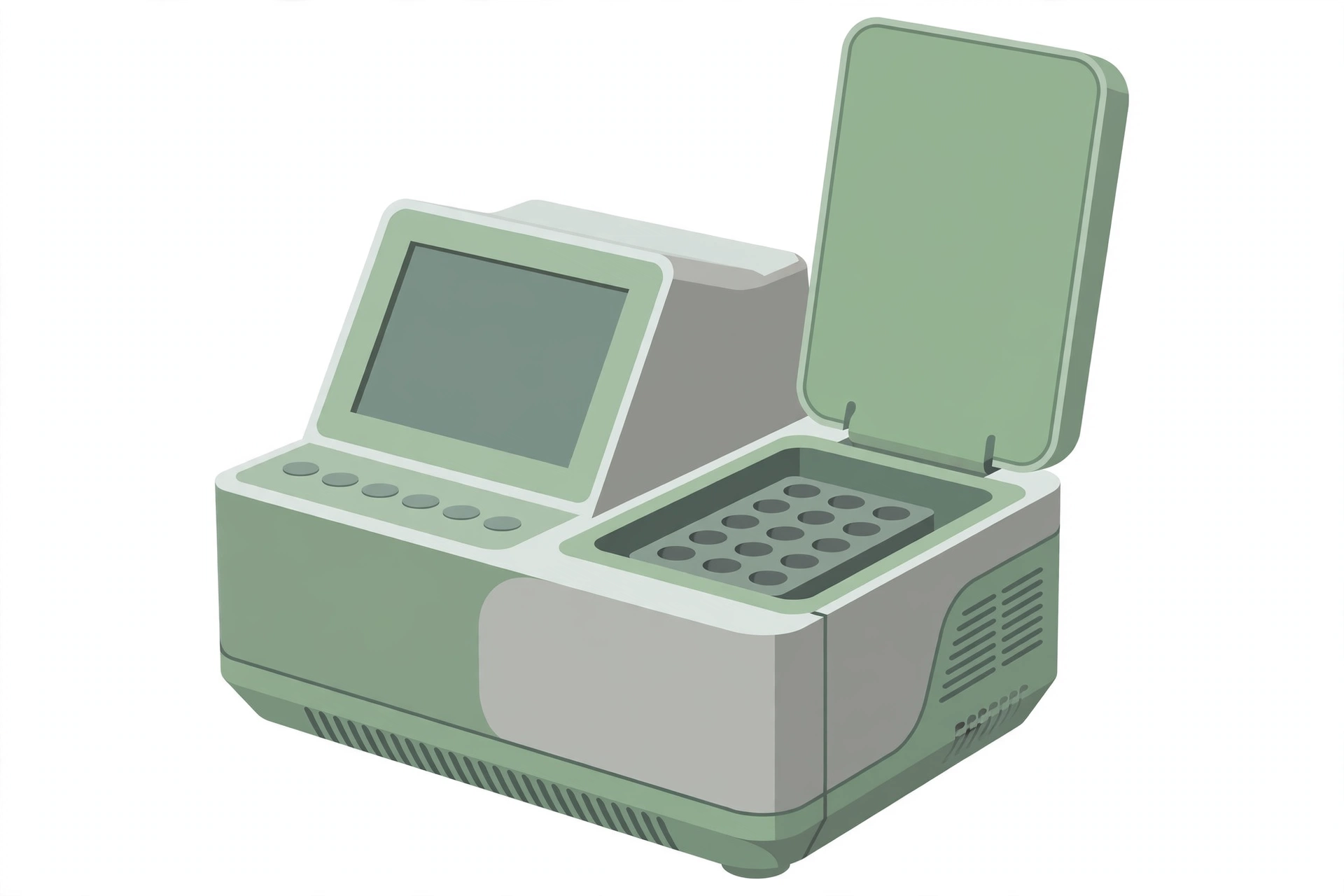
- AES-validated real-time PCR thermocycler with the following specifications:
- 96-well (or 48-well) microplate (low and high profile) or 96 (48) × 0.2 mL tubes (low and high profile) sample capacity
- the ability to excite fluorophores with a peak excitation range of 485 to 520 nm
- the ability to detect fluorophores with a peak emission range of 500 to 600 nm
- Conventional thermocycler for DNA extraction (optional)
- Centrifuge with rotor and microplate carriers
- Vortex equipped for microplates
- Two multi-channel pipettors
- Single-channel pipettor
- PCR enclosure (optional)
- PCR capping tool
- Strip holders
- ADIAFOOD Extraction kit (containing extraction plates, domed caps and seals)
- Aerosol barrier tips for pipettors
- Disposable multi-channel pipettor basins
- Sterile microcentrifuge screw cap tubes (2 mL)
- Powder-free gloves
- Required enrichment media
- Stomacher filter bags
- Disposable serological pipettes
- Alcohol (denatured) and bleach
- 0.1% peptone water
Reagents
- The oligonucleotide primers and molecular beacon probes are supplied in the kit and are specific for L. monocytogenes.
- The temperature cycling programs for the PCR include an automated extraction cycle, which lyses the bacterial cells, followed by an automated PCR detection process.
- All buffers and reagents are provided by AES Chemunex with each kit, including the extraction reagents (EX-1, EX-2) and the detection reagents (DT-1, DT-2). All other PCR components are pre-dispensed in the wells of the detection microplate or strips.
- All water, pipettes, pipette tips and other materials coming in contact with samples or PCR reagents should be sterile and DNase-free and/or autoclaved prior to use, to remove any DNase and/or other contamination.
Sample preparation
- Follow the handling of sample units, preparation for analysis, and preparation of samples described in the cultural method MFHPB-30.
Procedure
Pre-enrich samples in LEB for 24 h at 30 °C
↓
Transfer 200 µL of the LEB pre-enrichment to a Palcam agar slant and incubate at 35 °C for 18 to 24 hours
↓
Wash the agar slant with 2 mL of sterile peptone water to resuspend the cells; the resulting cell suspension is used for DNA extraction
↓
Refrigerate the remaining cell suspension
DNA extraction:
Dispense the contents from EX-1 (squeeze bottle) into the EX-2 vial to obtain reconstituted EX-2
↓
Recap the EX-2 vial and mix by inversion to ensure complete dissolution of the desiccated reagent
↓
Pour the reconstituted EX-2 into a multi-channel pipettor basin
↓
Aliquot 90 µL of reconstituted EX-2 into each well of the Extraction microplate
↓
Transfer 10 µL of the cell suspension into the Extraction microplate according to the sample assignment layout
↓
Seal the wells of the Extraction microplate with the domed cap strips provided
↓
Place the Extraction microplate into the thermocycler and begin the DNA extraction program on the thermocycler
↓
When the Extraction program is completed, take out the Extraction microplate; all organisms are now lysed and inactivated
↓
Centrifuge the Extraction microplate for 5 minutes at 1,800 × g; the supernatant can now be used in the PCR amplification method
PCR amplification:
Dispense the contents from DT-1 (squeeze bottle) into the DT-2 vial to obtain reconstituted DT-2
↓
Recap the DT-2 vial and mix by inversion to ensure complete dissolution of the desiccated reagent
↓
Pour the reconstituted DT-2 into a multi-channel pipettor basin
↓
Take out a Detection microplate or strips from its pouch (place them on the strip holder)
↓
Aliquot 15 µL of reconstituted DT-2 into the wells of the Detection microplate or strips
↓
Transfer 10 µL of extracted DNA from the Extraction microplate into the wells of the Detection microplate or strips
↓
Seal the Detection microplate or strips with the optical-grade caps provided
↓
Seal the Extraction microplate using the provided microplate seals and store at 4 °C until the analysis is completed
↓
Vigorously invert the Detection microplate or strips 4 times to ensure that the contents are thoroughly mixed in the wells
↓
Gently tap the Detection microplate or strips to remove any air bubbles from the bottom of the wells
↓
Vortex for 1 minute at maximum speed
↓
Centrifuge the Detection microplate or strips for 1 minute at 1,800 × g to ensure that all the PCR mixture, including the sample’s DNA extract, is in the bottom of the wells
↓
Secure the Detection microplate or strips tightly in the PCR instrument; make sure well A1 is in the upper left corner; click on “Start Detection” to initialize the PCR protocol
↓
When the Detection program is completed, remove and discard the Detection microplate or strips
Interpretation of results
- A positive PCR test results in a fluorescence response higher than the baseline produced by the negative controls within 40 cycles of the PCR amplification; the baseline cut-off fluorescence value is determined and set by the system.
- A negative PCR test normally does not produce visible fluorescence.
- If fluorescence occurs above the baseline cut-off value in the negative controls, the results of the test are invalidated and the analysis must be repeated with precautions taken to eliminate possible sources of error.
Confirmation of results
- Using the refrigerated cell suspension, proceed with the plating and confirmation steps of a cultural method — MFHPB-07 or MFHPB-30.
Precautions
- The laboratory supervisor must ensure that completion of the analysis described in this method is done in accordance with the International Standards reference “ISO/IEC 17025:1999 (or latest version): General Requirements for the Competence of Testing and Calibration Laboratories”.
- All water, pipettes, pipette tips and other materials coming in contact with samples or PCR reagents should be sterile and DNase-free and/or autoclaved prior to use.
References
- Health Canada. Identification of Listeria monocytogenes using the Adiafood Rapid Pathogen Detection System, a Real-Time PCR Technique. Compendium of Analytical Methods, Volume 3, Laboratory Procedure MFLP-31, October 2011. Bureau of Microbial Hazards, Food Directorate, Health Products and Food Branch, Health Canada.
- Pagotto, F., E. Daley, J. Farber and D. Warburton. 2001. MFHPB-30. Isolation of Listeria monocytogenes from all Foods and Environmental Samples. In: Volume 2, Compendium of Analytical Methods.
- Warnex Research Inc. 2002. Validation of the Listeria monocytogenes Real Time PCR Technology for the Rapid Identification of Listeria monocytogenes from Food and other Samples. Personal Communication (unpublished data).
- AdiaFood user manual published by AES Chemunex, including the product insert for the enrichment and detection of Listeria monocytogenes.