SOP: Enumeration of generic E. coli and other coliform bacteria from all foods using RAPID'E. coli 2 chromogenic media
GIFSQ — Global Institute for Food Safety & Quality
Principle
This method is applicable to the rapid detection of generic Escherichia coli and other coliform bacteria, to determine compliance with the requirements of Sections 4 and 7 of the Food and Drugs Act and/or other relevant federal regulations. The method has been validated for the enumeration of coliforms and E. coli from all foods.
Coliforms are a non-taxonomic group of Gram-negative, non-spore-forming, facultative anaerobic, rod-shaped bacteria that ferment lactose to produce acid and gas. The group includes enteric bacteria such as Escherichia, Citrobacter, Klebsiella and Enterobacter. Enumeration of coliform bacteria can be useful to confirm the sanitation of water or food-processing environments. E. coli is found in the intestines of animals and is abundant in human and animal feces; it can also be isolated from environmental samples. Fecal coliforms, used as indicators of contamination and the possible presence of pathogenic organisms, consist primarily of E. coli and some thermotolerant Klebsiella.
RAPID’E. coli 2 medium relies on the simultaneous detection of two enzymatic activities: β-D-glucuronidase (GLUC) and β-D-galactosidase (GAL). The medium contains two chromogenic substrates. One substrate is specific to GAL and results in blue coloration of colonies positive for this enzyme, and one substrate is specific to GLUC and results in violet coloration of colonies positive for this enzyme. Coliforms other than E. coli (GAL+/GLUC−) form blue to green colonies, while E. coli (GAL+/GLUC+) form violet to pink colonies. A count of total coliforms is obtained by adding the number of blue-green colonies and the number of violet to pink colonies.
Apparatus
- Balance — for weighing food samples
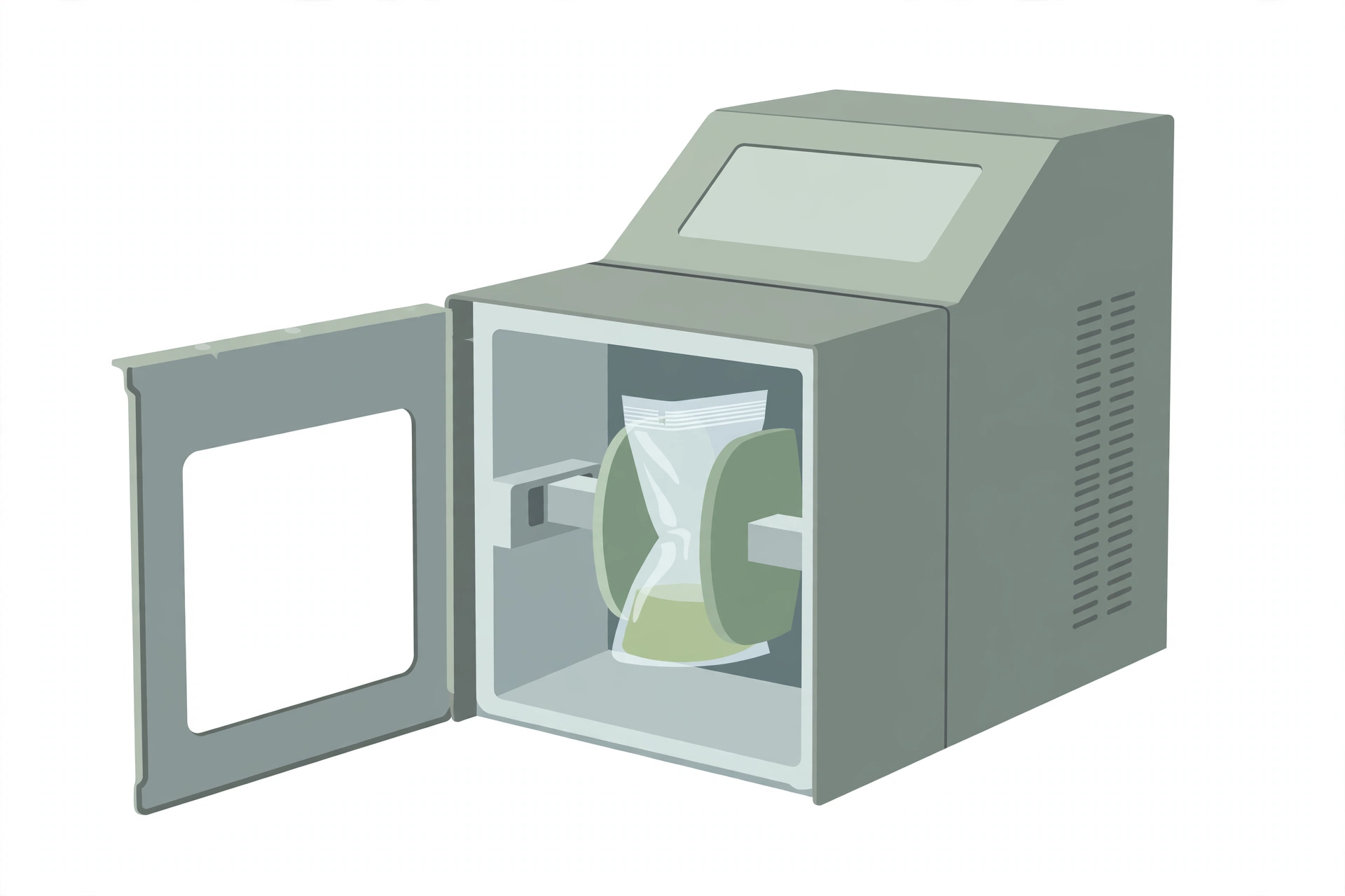
- Stomacher, blender, vortex or equivalent
- Incubator capable of maintaining temperatures of 37 ± 1 °C and 44 ± 0.5 °C
- Water bath capable of maintaining temperatures of 44 – 47 °C
Reagents
- RAPID’E. coli 2 agar:
- Ready-to-use 100 mL x 6 bottles (Bio-Rad catalog # 3555299)
- Ready-to-use 200 mL x 6 bottles (Bio-Rad catalog # 3555297)
- Dehydrated medium 500g (Bio-Rad catalog # 3564024)
- Butterfield’s phosphate buffer
- Stomacher bag, or stomacher bag with incorporated filter (recommended for meat and highly particulate samples)
- Sterile pipettes
- Sterile petri dishes
- Brilliant Green Lactose Bile broth (BGLB)
- Levine’s Eosin Methylene Blue agar (L-EMB)
- Lauryl Sulfate Tryptose (LST) broth
- (EC) E. coli broth
Media formulations
- Butterfield’s Phosphate Buffer
- Stock Solution
- Potassium phosphate monobasic (KH₂PO₄) — 34.0 g
- Distilled water — 500 mL
- pH to 7.2
- Complete Medium: adjust the pH to 7.2 with 1 N NaOH and bring the volume to 1.0 L with distilled water; sterilize at 121 °C for 15 min and store in the refrigerator
- Dilution blanks: take 1.25 mL of the stock solution and bring the volume to 1.0 L with distilled water, dispense the required volume and sterilize at 121 °C for 15 min
- Brilliant Green Lactose Bile (BGLB) Broth
- Basal Medium
- Peptone — 10.0 g
- Lactose — 10.0 g
- Ox-gall — 20.0 g
- Brilliant green — 0.0133 g
- Final pH 7.2 ± 0.2
- Complete Medium: dissolve the peptone and lactose in 500 mL of water; add the ox-gall dissolved in 200 mL of water; bring the volume to approximately 975 mL with water and adjust the pH; add 13.3 mL of a 1% aqueous solution of brilliant green; bring the total volume to 1.0 L, stir, and filter through cotton if necessary to clarify; dispense in 10.0 mL volumes into tubes containing gas vials; sterilize at 121°C for 12 min
- Levine’s Eosin Methylene Blue (L-EMB) Agar
- Basal Medium
- Peptone — 10.0 g
- Lactose — 10.0 g
- Dipotassium phosphate (K₂HPO₄) — 2.0 g
- Agar — 15.0 g
- Eosin Y — 0.4 g
- Methylene blue — 0.065 g
- Distilled water — 1.0 L
- Final pH: 7.1 ± 0.2
- Complete Medium: add all the ingredients to distilled water; heat to boiling to obtain a complete solution; mix well; dispense as required (usually in 100–200 mL volumes); sterilize at 121 °C for 15 min; avoid overheating, because the agar is likely to become soft and unsuitable for streaking
- Lauryl Sulfate Tryptose (LST) Broth
- Basal Medium
- Tryptose, tryptone or trypticase — 20.0 g
- Lactose — 5.0 g
- Dipotassium phosphate (K₂HPO₄) — 2.75 g
- Monopotassium phosphate (KH₂PO₄) — 2.75 g
- Sodium chloride (NaCl) — 5.0 g
- Sodium lauryl sulphate — 0.1 g
- Distilled water — 1.0 L
- Final pH: 6.8 ± 0.2
- Complete Medium: dissolve the ingredients in 1.0 L of distilled water (in 500 mL if double strength is required); dispense in 10.0 mL volumes into tubes containing gas vials; sterilize at 121 °C for 15 min
- EC (E. coli) Broth
- Basal Medium
- Tryptose or trypticase — 20.0 g
- Lactose — 5.0 g
- Bile salts No. 3 — 1.5 g
- Di-potassium phosphate (K₂HPO₄) — 4.0 g
- Mono-potassium phosphate (KH₂PO₄) — 1.5 g
- Sodium chloride (NaCl) — 5.0 g
- Distilled water — 1.0 L
- pH 6.9 ± 0.2
- Complete Medium: dissolve the ingredients in 1.0 L of distilled water, heating gently if necessary to obtain a complete solution; dispense in 10.0 mL volumes into tubes containing gas vials; sterilize at 121 °C for 15 min
Sample preparation
Handling of sample units
- Before analysis in the laboratory, except for shelf-stable foods, keep sample units refrigerated or frozen, depending on the nature of the product.
- Thaw frozen samples in a refrigerator, or under time and temperature conditions which prevent microbial growth or death.
- Analyze sample units as soon as possible after their receipt in the laboratory.
Preparation for analysis
- Have sterile Butterfield’s phosphate buffer diluent prepared.
- Clean the surface of the working area with a suitable disinfectant.
Preparation of sample
- To ensure a representative analytical unit, agitate liquids or free-flowing materials until the contents are homogeneous.
- If the sample unit is a solid, obtain the analytical unit by taking a portion from several locations within the sample unit.
- Prepare a 1 in 10 dilution of the food by aseptically adding 11 (50) g or mL of the food to 99 (450) g or mL of sterile Butterfield’s phosphate buffer diluent (weight or volume in brackets indicates the alternate procedure for making dilutions).
- Blend, stomach or vortex as required for thorough mixing.
- Prepare serial dilutions as necessary in sterile Butterfield’s phosphate buffer diluent.
Procedure
Each sample unit shall be analyzed individually.
Plating
- Agitate each dilution to resuspend material that may have settled during preparation. Plating should be carried out rapidly after preparing the dilutions
↓ - Using a sterile pipette, transfer 1 mL of the sample to be tested and/or 1 mL of its serial dilutions to a sterile petri dish. Samples should be plated in duplicate
↓ - Pour 15–20 mL of agar, tempered to 44 – 47 °C, into the petri dish directly on top of the sample. The plate should be poured not more than 15 min after the dilution preparation
↓ - Swirl the petri dish to homogenize the sample and agar
↓ - Allow the plate to solidify
Incubation
- Incubate the plate in the inverted position at:
- 37 °C for 18 – 24 h for simultaneous enumeration of E. coli and coliforms (= total coliforms)
- AND/OR
- 44 °C for 18 – 24h for enumeration of E. coli
↓ - It is recommended to incubate in stacks of no more than three plates
↓ - Avoid excessive crowding of plates in order to permit rapid equilibration of the plates with incubator temperature
Counting colonies
- After incubation, proceed with counting typical colonies on the plate
↓ - Count all blue to green colonies as coliform bacteria (other than E. coli). If required, proceed with confirmation of presumptive positive coliforms
↓ - Count all violet to pink colonies as E. coli. If required, proceed with confirmation of presumptive positive E. coli
↓ - Proceed to Calculation for the enumeration calculations
Confirmation (optional)
- For non-E. coli coliforms: select two suspect non-E. coli coliform colonies and inoculate two tubes containing Lauryl Sulfate Tryptose (LST) broth. Incubate the tubes at 35 °C and examine for gas formation at 24 and 48 h
↓ - Using an inoculating loop, transfer a loopful of broth from the gassing tubes to Brilliant Green Lactose Bile broth (BGLB broth) and incubate at 35 °C for 48 ± 2 h
↓ - Colonies producing growth and gas in LST and BGLB are considered confirmed for non-E. coli coliform bacteria. Determine the number of non-E. coli coliforms by multiplying the percent of colonies confirmed as positive by the original RAPID’E. coli 2 count. Multiply the confirmed number of colonies by the appropriate dilution factor
↓ - For E. coli coliforms: select two suspect E. coli colonies and inoculate two tubes containing LST broth. Incubate the tubes at 35 °C and examine for gas formation at 24 and 48 h
↓ - Using an inoculating loop, transfer a loopful of broth from the gassing tubes to (EC) E. coli broth and incubate at 45.5 °C for 48 ± 2 h
↓ - Streak a loopful of broth from the gas-positive tubes onto Levine’s Eosin Methylene Blue agar (L-EMB) and incubate at 35 °C for 24 ± 2 h. Select a minimum of 2 typical colonies with dark purple center and green metallic sheen from each L-EMB plate for confirmation. For each colony selected, perform a Gram stain and inoculate an additional tube of LST broth — incubate at 35 °C for 48 ± 2 h, and examine for gas formation. Conduct additional biochemical tests as follows: indole, methyl red, Vogues-Proskauer, and citrate tests. These biochemical tests may be performed using a rapid ID kit that encompasses the tests specified above
↓ - Confirmed E. coli isolates are Gram-negative, exhibit growth and gas in LST, are indole and methyl red positive, and Vogues-Proskauer and citrate negative
↓ - Determine the total number of E. coli colonies by multiplying the percent of isolates confirmed as positive by the original RAPID’E. coli 2 E. coli count. Multiply the confirmed number of E. coli by the appropriate dilution factor. Since E. coli is a coliform bacterium, enumeration of total coliforms is achieved by adding the number non-E. coli coliforms to the number of E. coli
Calculation
E. coli only CFU/g or mL = All violet to pink colonies × dilution factor
Other coliforms CFU/g or mL = All blue to green colonies × dilution factor
Total coliform CFU/g or mL = All blue to green and violet to pink colonies × dilution factor
Where,
- All violet to pink colonies = the number of violet-to-pink colonies counted on the selected dishes (all E. coli colonies)
- All blue to green colonies = the number of blue-to-green colonies counted on the selected dishes (all non-E. coli coliform colonies)
- dilution factor = the appropriate dilution factor for the dilution plated
Recording results
- To achieve enumeration of the total number of coliform bacteria, add the number of blue to green and violet to pink colonies and multiply by the appropriate dilution factor.
- Select only dishes containing fewer than 300 total coliform colonies in all.
- Calculate the number of CFU per g or mL by multiplying the number of colonies present by the appropriate dilution factor.
- Report the actual count whenever possible. If no colonies are recovered in the 1 in 10 dilution, report as < 10 CFU/g or mL. If more than 300 colonies in all appear, the food sample may be retested using a higher dilution (i.e., 1 in 50 or 1 in 100) to enable a more accurate enumeration, or reported as > 300 × the dilution factor.
Precautions
- The laboratory supervisor must ensure that completion of the analysis described in this method is done in accordance with the International Standards reference “ISO/IEC 17025:2005 (or latest version): General Requirements for the Competence of Testing and Calibration Laboratories”.
- It is the responsibility of the laboratory to ensure equivalency if any variations of the media formulations listed here are used (either product that is commercially available or made from scratch).
- It is the responsibility of each laboratory to ensure that the temperatures of the incubators or water baths are maintained at the recommended temperatures. Where 35 °C is recommended in text of the method the incubator may be at 35 ± 1.0 °C. Similarly, lower temperatures of 30 or 25 may be ± 1.0 °C. However, where higher temperatures are recommended, such as 43 or 45.5 °C, it is imperative that the incubators or water baths be maintained within 0.5 °C due to potential lethality of the higher temperatures on the microorganism(s) being isolated.
References
- Health Canada, Health Products and Food Branch. Enumeration of generic E. coli and other coliform bacteria from all foods using RAPID’E. coli 2 chromogenic media. Compendium of Analytical Methods, Volume 3, Laboratory Procedure MFLP-64, May 2018.
- Bio-Rad Laboratories – RAPID’E. coli 2 agar Technical Sheet.
- Official Methods of Analysis (1995) 16th Ed., AOAC INTERNATIONAL, Gaithersburg, MD, Method 966.24.