SOP: Enumeration of Escherichia coli in Foods by a Direct Plating (DP) Method
GIFSQ — Global Institute for Food Safety & Quality
1. Principle
Escherichia coli biotype 1 is enumerated in foods and food ingredients to determine compliance with the requirements of Sections 4 and 7 of the Food and Drugs Act. The conventional Most Probable Number (MPN) procedure takes 8 to 12 days to complete; the Direct Plating (DP) method takes 24 to 30 h. In previous studies comparing the two, the MPN procedure proved less precise and gave lower E. coli counts in frozen meat samples, and it cannot enumerate late lactose fermenters or anaerogenic E. coli, which make up approximately 10% of the E. coli strains. The main drawback of the DP method is that it cannot enumerate non-indole producing E. coli (3 to 5% of the E. coli strains); this is offset by how fast the method is and by its facility in enumerating anaerogenic E. coli.
The DP method has been used successfully for red meats, poultry, pork, sausages, carcass washings, fish, shellfish, milk, ice cream, cheese, apple and strawberry purées, flour, carrots, potatoes, swede, and green beans. It is not applicable to bean and alfalfa sprouts, where large numbers of Klebsiella spp. may preclude an accurate determination of E. coli.
2. Apparatus required
- Incubator capable of maintaining 44.5 ± 0.5 °C
- Incubator capable of maintaining 35 ± 1 °C
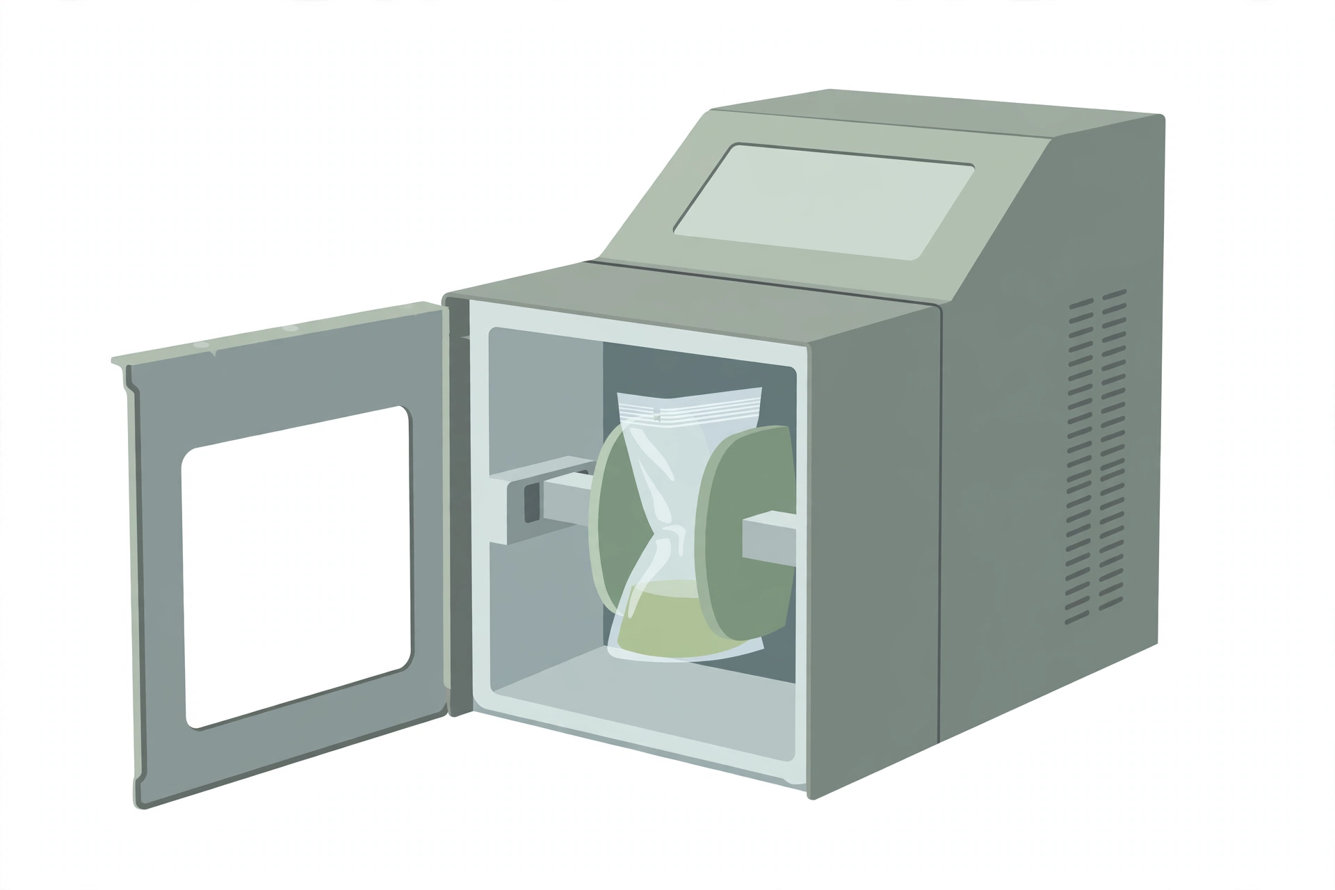
- Stomacher, blender or equivalent
- Membrane filter forceps
- Sterile glass or plastic spreader
- Petri plates (for duplicate plating)
- ‘Germicidal’ UV lamp (Philips TUV, 15 W or GE G 30T8, 30 W)
- Fume hood or biocontainment unit (for the indole-staining/UV step)
3. Materials and reagents
- 0.45 µm membrane filters, 85 mm diameter (Cat. No. CA87003-868, VWR)
- Peptone water (0.1%)
- To prepare: suspend 1.0 g of peptone in 1.0 L of distilled water; mix thoroughly and dispense; autoclave at 121 °C for 15 min. Final pH 7.0 ± 0.2.
- Nutrient agar (NA), prepoured and dried
- Basal medium: peptone — 5.0 g; beef extract — 3.0 g; agar — 15.0 g; distilled water — 1.0 L. pH 6.8 ± 0.2.
- To prepare: suspend the ingredients in 1.0 L of distilled water and heat to boiling with frequent swirling ↓ dispense into tubes or plates and autoclave at 121 °C for 15 min. Cool tubes in a slanted position. Plates can be stored for up to 8 weeks at 4 °C.
- Tryptone Bile Agar (TBA), prepoured and dried
- Basal medium: tryptone — 20.0 g; bile salts no. 3 — 1.5 g; agar — 15 g; distilled water — 1.0 L. pH to 7.2 ± 0.2.
- To prepare: suspend the basal medium ingredients in 1.0 L of distilled water ↓ heat to boiling with constant stirring to dissolve all ingredients ↓ autoclave at 121 °C for 15 min ↓ pour 15–20 mL per plate on a flat surface.
- Indole reagent
- Suitable disinfectant (for the working area)
- Positive and negative control cultures, ATCC or equivalent
4. Sample preparation
Analyze each sample unit individually. Except for shelf-stable foods, keep the sample units refrigerated or frozen, depending on the nature of the product. Thaw frozen samples in a refrigerator, or under time and temperature conditions which prevent microbial growth or death. Analyze the sample units as soon as possible after receipt at the laboratory.
- Clean the surface of the working area with a suitable disinfectant.
- Label duplicate Petri plates with the appropriate identifying information.
- Have sterile peptone dilution water ready.
- Agitate liquids or free-flowing materials until the contents are homogeneous. For a solid sample unit, obtain the analytical unit by taking a portion from several locations within the unit.
- For solid samples, blend or stomach 11 (10) g (the analytical unit) with 99 (90) mL of peptone water for 2 min, then make decimal dilutions in peptone water. If a low count is expected, blend 11 (10) g with 44 (40) mL of peptone water (1:5).
- For liquid samples, shake 11 (10) mL with either 99 (90) mL (1:10) or 44 (40) mL (1:5) of peptone water. Liquid samples may also be plated undiluted.
- The weight or volume in brackets indicates the alternative procedure for making dilutions.
5. Procedure
If the organisms in the sample may have been stressed by freezing or other processing, follow the NA pre-incubation path; otherwise, go directly to the TBA path.
Stressed samples — place an MF on the dried surface of a prepoured NA plate and flatten it with a sterile glass or plastic spreader, trapping no air bubbles
↓
In duplicate, plate 0.5 mL of two consecutive decimal dilutions — or, if low counts are expected, 1 mL of the 1:5 dilution, or 0.5 mL of the undiluted sample (liquid samples) — onto the MF overlaying the NA, and spread the inoculum evenly with a glass or plastic spreader without spilling it over the edge of the MF
↓
After the inoculum has been absorbed, incubate the plates right side up, in stacks of not more than three deep, in a 35 ± 1 °C incubator for 4 h
↓
Remove the MF from the NA with sterile forceps and transfer it to the dried surface of a prepoured TBA plate, trapping no air bubbles
↓
Incubate the TBA plates right side up, in stacks of not more than three deep, in a 44.5 ± 0.5 °C incubator for 18–24 h
Unstressed samples (the NA step is omitted) — place an MF on the dried surface of a prepoured TBA plate and flatten it with a sterile glass or plastic spreader, trapping no bubbles
↓
In duplicate, plate onto the MF overlaying the TBA exactly as above, and incubate right side up, in stacks of not more than three deep, at 44.5 ± 0.5 °C for 20–24 h
After the 44.5 ± 0.5 °C incubation, remove the Petri plate covers, wipe them dry, and place 2.0 mL of indole reagent into each cover
↓
Lift each MF off the TBA surface and place it into its respective cover so the entire undersurface of the MF is soaked with the reagent; leave at room temperature for 10–15 minutes
↓
Remove the MF by dragging it over the lip of the cover to get rid of excess reagent, and place it on a flat surface (e.g. another clean Petri plate cover) under a ‘germicidal’ UV lamp for approximately 20 minutes — this is best done in a fume hood or biocontainment unit, to avoid exposure to the HCl fumes given off
↓
The pink to red colonies appearing on the MF are indole producers; enumerate them as E. coli biotype I
- If colonies are to be kept for any reason, pick them from the MF before staining.
- Use the dilution factor to calculate the number of E. coli biotype I per g or mL, and record the result.
6. Calculation
Number of CFU per g or mL = (number of CFU) ÷ [volume plated (mL) × total dilution factor]
Where,
- number of CFU — the colonies counted on the filters (duplicate filters are summed)
- volume plated (mL) — the volume spread on each filter
- total dilution factor — the dilution factor of the suspension plated (e.g. 0.2 for a 1:5 dilution)
Worked example (as the method gives it):
- A 1:5 dilution is made from a food matrix. 0.5 mL is spread onto each of two MFs overlaying TBA plates. After incubation, one MF has 35 colonies and the other has 41 colonies.
- (35 + 41) ÷ [1 (volume plated, mL) × 0. 2 (dilution factor)] = 380 CFU/g
Reporting notes:
- Report the actual count whenever possible.
- If there are no colonies in the 1:5 dilution, report as < 5 CFU/g. If no colonies are recovered in the 1:10 dilution, report as < 10 CFU/g.
- If more than 200 typical colonies appear, retest the food sample at a higher dilution (1:50 or 1:100) to enable a more accurate enumeration.
7. Precautions
- The laboratory supervisor must ensure that the analysis is carried out in accordance with ISO/IEC 17025:1999 (or the latest version), General Requirements for the Competence of Testing and Calibration Laboratories.
- It is the responsibility of the laboratory to ensure equivalency if any variations of the listed media formulations are used, whether commercially available or made from scratch; forward equivalency data to the Editor of the Compendium of Analytical Methods for consideration of a modification to this method.
- Incubator and water-bath temperatures must be maintained at the recommended temperatures. Where 35 °C is recommended, the incubator may be at 35 ± 1.0 °C; lower temperatures of 30 or 25 °C may be ± 1.0 °C. Where higher temperatures are recommended, such as 43 or 45.5 °C, the incubators or water baths must be held within 0.5 °C, because of the potential lethality of the higher temperatures on the microorganism being isolated.
- The method’s UV step is printed as “approximatley 20 minutes”.
References
Health Canada, Compendium of Analytical Methods, Volume 2 — HPB Method MFHPB-27, October 2012: “Enumeration of Escherichia coli in Foods by a Direct Plating (DP) Method.” (This revised method replaces MFHPB-27, September 1997.)
- Anderson, J.M., and A.C. Baird-Parker. 1975. A rapid and direct plate method for enumerating Escherichia coli biotype I in food. J. Appl. Bacteriol. 39: 111–117.
- Ewing, W.H. 1972. Differentiation of enterobacteriaceae by biochemical reactions. CDC Atlanta, U.S. Dept. of Health, Education and Welfare.
- Holbrook, R., J.M. Anderson, and A.C. Baird-Parker. 1980. Modified direct plate method for counting Escherichia coli in foods. Food Technol (Ans.) 32: 78–83.
- Rayman, M.K., and B. Aris. 1981. The Anderson-Baird-Parker direct plating method versus the most probable number procedure for enumerating Escherichia coli in meats. Can. J. Microbiol. 27: 147–149.
- Rayman, M.K., G.A. Jarvis, C.M. Davidson, S. Long, J.M. Allen, T. Tong, P. Dodsworth, S. McLaughlin, S. Greenberg, B.G. Shaw, H.J. Beckers, S. Qvist, P.M. Nottingham, and B.J. Stewart. 1979. ICMSF methods studies. XIII. An international comparative study of the MPN procedure and the Anderson-Baird-Parker direct plating method for the enumeration of Escherichia coli biotype I in raw meats. Can. J. Microbiol. 25: 1321–1327.
- Sharpe, A.N., P.I. Peterkin, and M.K. Rayman. 1981. Detection of Escherichia coli in foods: Indole staining methods for cellulosic and polysulfone membrane filters. Appl. Environ. Microbiol. 41: 1310–1315.
- Sharpe, A.N., M.K. Rayman, D.M. Burgener, D. Conley, A. Loit, M. Milling, P.I. Peterkin, U. Purvis, and S. Malcolm. 1983. Collaborative study of the MPN, Anderson/Baird-Parker direct plating, and hydrophobic grid-membrane filter methods for the enumeration of Escherichia coli in foods. Can. J. Microbiol. 29: 1247–1252.
- Yoovidhya, T., and G.H. Fleet. 1981. An evaluation of the A-1 most probable number and the Anderson and Baird-Parker plate count methods for enumerating Escherichia coli in the Sydney rock oyster, Crassostrea commercialis. J. Appl. Bacteriol. 50: 519–528.