SOP: The Dupont Qualicon Bax System Method for the Detection of Enterobacter sakazakii in Selected Foods
GIFSQ — Global Institute for Food Safety & Quality
Principle
This method is applicable to the detection of Enterobacter sakazakii in selected foods, including powdered infant formula, dry dairy and soy ingredients, and food production environmental samples after enrichment.
The BAX system is a yes/no screening tool that uses Polymerase Chain Reaction (PCR) technology for rapid amplification and fluorescent detection, giving a quick and accurate way to detect Enterobacter sakazakii in a wide variety of foods. After 20–22 hours of primary enrichment and 3 hours of secondary enrichment, sample preparation involves about 1 hour of user time, and the automated procedure delivers reliable results about 4 hours later. The BAX system validation studies used an enrichment method developed by the Nestle Research Centre. The BAX system is designed for use by qualified laboratory personnel who follow standard microbiology practices.
The BAX system uses PCR to amplify a specific fragment of bacterial DNA that is stable and unaffected by the growth environment. The fragment is a genetic sequence unique to Enterobacter sakazakii, providing a highly reliable indicator that the organism is present; the automated BAX system then uses fluorescent detection to analyze the PCR product for positive or negative results. PCR rapidly provides millions of copies of a specific DNA fragment: sample DNA is combined with polymerase, nucleotides and primers specific for a given nucleotide sequence, and the mixture undergoes a series of timed heating and cooling cycles. Heating denatures the DNA into single strands; as the mixture cools, the primers recognize and anneal to the target DNA sequence, and DNA polymerase extends the primers using the nucleotides, creating two copies of the target DNA fragment. Repeated cycles of denaturing, annealing and extending produce exponential increases in the number of target DNA fragments within hours. If the target sequence is not present, no detectable amplification takes place. The BAX system simplifies this process by combining the primers, polymerase, nucleotides and positive control into a single sample tablet already packaged inside the PCR tubes; automated fluorescent detection allows closed-tube testing, eliminating the potential for carry-over contamination with amplified DNA.
Apparatus
Equipment
- Stomacher
- Incubator
- Other (included with BAX system start-up package):
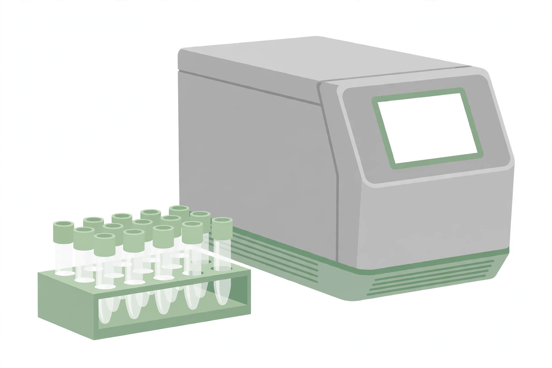
- Cycler/detector with verification plates
- Computer workstation with Microsoft Windows operating system, BAX system application, and printer
- Dry block heaters with thermometers inserts for lysis tubes
- Capping/decapping tools
- Various pipettes for reagent and sample transfers
- Cooling block with inserts for lysis tubes and PCR tubes
- PCR tube holders
Supplies
- Lysis tubes with caps and rack
- Tips for pipettes
- Powder-free nitrile gloves
- BAX system User Guide
Reagents
- BAX system kit no. 17720657 (sufficient for 96 tests; DuPont Qualicon), containing:
- 2 bags of PCR sample tablets — 1 tablet packaged per PCR tube in 12 strips of 8 tubes; the tablets include the reagents needed for the test reaction as well as an internal positive control (which eliminates the need to run a separate QC reaction); tablets are 7.6 ± 0.1 mg
- 1 bag of optical caps — 12 strips of 8 caps
- 2 bottles of ES lysis buffer — pH 8.35 ± 0.05 at 25 °C; 12 mL/bottle; used to prepare the working lysis reagent
- 1 vial of protease solution — 400 µL/vial; used to prepare the working lysis reagent
- Primary enrichment — mLST broth with vancomycin (prepared as below)
- Secondary enrichment — BHI broth (commercially available)
- To prepare: basal medium — infusion from 200.0 g calf brain, infusion from 250.0 g beef heart, 10.0 g proteose peptone, 2.0 g dextrose, 5.0 g sodium chloride (NaCl) and 2.5 g sodium phosphate (Na₂HPO₄) per 1.0 L of distilled water; final pH 7.4 ± 0.2. Dissolve the ingredients in 1.0 L of distilled water, distribute as required (tubes or flasks), and sterilize at 121 °C for 15 min.
- Confirmation medium — nutrient agar (commercially available)
- To prepare: suspend 5.0 g peptone, 3.0 g beef extract and 15.0 g agar in 1.0 L of distilled water (pH 6.8 ± 0.2); heat to boiling with frequent swirling; dispense into tubes or plates and autoclave at 121 °C for 15 min. Cool the tubes in a slanted position. Plates can be stored for up to 8 weeks at 4 °C.
- Alpha-glucosidase tablets (Rosco Diatab 50421)
Preparation of mLST broth with vancomycin
- Mix 20 g tryptose, 5 g lactose, 2.75 g K₂HPO₄, 2.75 g KH₂PO₄, 34.22 g NaCl and 0.1 g sodium lauryl sulfate in 1 liter of distilled water; autoclave at 121 °C for 15 minutes
- Prepare vancomycin as a 10 mg/mL solution in distilled water; filter-sterilize through a 0.2 µm filter, then transfer 1 mL of the vancomycin solution to 1 liter of the cooled (less than 50 °C) modified LST broth
- Use this broth within 24 hours
- Commercial brands of LST media may also be used, but extra NaCl (29.22 g/liter) must be added to the broth before autoclaving
- To prepare Lauryl Sulfate Tryptose (LST) broth from scratch: dissolve 20.0 g of tryptose, tryptone or trypticase, 5.0 g of lactose, 2.75 g of dipotassium phosphate (K₂HPO₄), 2.75 g of monopotassium phosphate (KH₂PO₄), 5.0 g of sodium chloride (NaCl) and 0.1 g of sodium lauryl sulphate in 1.0 L of distilled water (in 500 mL if double strength is required); final pH 6.8 ± 0.2. With the stated modification, add the extra 29.22 g of NaCl per litre (34.22 g total) before autoclaving. Dispense in 10.0 mL volumes into tubes containing gas vials and sterilize at 121 °C for 15 min.
Procedure
Blend 25 g of sample with 225 mL of mLST broth with vancomycin
↓
Incubate at 45 °C for 20–22 hours
↓
Add 10 µL of the enriched sample to 500 µL of BHI broth (room temperature)
↓
Incubate at 37 °C for 3 hours
↓
Make sure the cooling blocks have been refrigerated overnight
↓
Warm up the heating blocks; check that the temperatures are set to 37 °C and 95 °C
↓
Initialize the BAX system cycler/detector (perform verification, if prompted)
↓
Create a rack file
↓
Select RUN FULL PROCESS in the menu bar to warm up the cycler/detector
↓
Arrange the required number of lysis tubes (one for each sample and one for the “blank”) in the rack according to the rack file
↓
Prepare the lysis reagent by pipetting 150 µL of protease into one 12-mL bottle of ES lysis buffer
↓
Add 200 µL of lysis reagent to each lysis tube
↓
Transfer 5 µL of enriched sample to the corresponding lysis tube
↓
Secure the caps and heat the tubes at 37 °C for 20 minutes, then at 95 °C for 10 minutes
↓
Place the lysis tubes in the cooling block for at least 5 minutes
↓
Place the PCR tube holder onto the PCR cooling block
↓
Arrange one PCR tube per sample in the holder
↓
Remove and discard the lid from one strip of tubes at a time
↓
Using a multi-channel pipette, transfer 50 µL of each lysed sample into a corresponding PCR tube
↓
Cover the tubes with a new optical cap strip and secure tightly; repeat for all samples
↓
Take the entire cooling block to the cycler/detector; keep the samples in the cooling block until the cycler/detector is ready for loading — no more than 30 minutes after tablet hydration
↓
Follow the screen prompts of the PCR Wizard to load the samples, run the program and unload the samples, as specified in the User Guide
↓
Review the results in the BAX system application — each sample is displayed as Negative (green circle with a minus sign), Positive (red circle with a plus sign), Indeterminate (yellow circle with a question mark) or Signal error (yellow and red circle with a question mark)
↓
Plate and streak from the original enrichments onto nutrient agar
↓
Incubate the nutrient agar plates at 37 ± 1 °C for 24 hours, then remove the plates from the incubator and place them under light for 4 hours at room temperature
↓
Pick up to 5 yellow colonies and perform the alpha-glucosidase test as follows: transfer 0.25 mL saline into a small glass tube
↓
Suspend 2–3 yellow colonies and one diagnostic tablet into the saline by vortexing
↓
Incubate in a water bath at 37 ± 1 °C for 4 hours
↓
Observe the colour of the liquid: bright yellow is a positive result
↓
Run one positive E. sakazakii control and one sterile saline negative control alongside the test
↓
If the colonies produce negative results, select up to 5 white colonies (atypical Enterobacter sakazakii) and perform the alpha-glucosidase test on them
↓
Positive results from the alpha-glucosidase test are considered presumptive and need further biochemical (API 20E) or molecular (RiboPrinter system) identification for confirmation as Enterobacter sakazakii
References
- Health Canada. The Dupont Qualicon Bax System Method for the Detection of Enterobacter sakazakii in Selected Foods. Compendium of Analytical Methods, Volume 3, Laboratory Procedure MFLP-27, September 2003. Health Products and Food Branch, Health Canada.