SOP: Isolation of Yersinia enterocolitica from Foods and Environmental Samples
GIFSQ — Global Institute for Food Safety & Quality
Principle
This method is applicable to the isolation and identification of Yersinia enterocolitica from foods and environmental samples to determine compliance with the requirements of Sections 4 and 7 of the Food and Drugs Act. This method replaces MFLP-48, dated January 1999.
Y. enterocolitica is a Gram-negative bacillus, from 1.0 to 3.5 µm in length and from 0.3 to 1.3 µm in width. When incubated at 25 °C, the vegetative cells in young cultures are ovoid to rod-shaped and later develop into the characteristic rod-shaped morphology. The cells are motile at 22–25 °C via peritrichous flagella, but are non-motile at 35–36 °C. The temperature range for growth is −2 to 40 °C with optimum growth at 18–30 °C for many strains. Serologically, 57 somatic “O” factors have been recognized, some of which are common to the genera Brucella, Vibrio and Salmonella. This method produces satisfactory results with naturally-contaminated food.
The method utilizes enrichment media and selective plating for the isolation of Yersinia enterocolitica from naturally contaminated food products. To detect small numbers of Yersinia in the presence of large numbers of contaminating microorganisms in food, a selective broth enrichment step should be carried out. There are two enrichment systems: one- and two-step enrichments. The one-step procedure has shown slightly lower recovery of certain strains of Y. enterocolitica than the two-step enrichment procedure; however, the one-step method requires a shorter incubation time and a simpler medium. For isolation of most pathogenic strains the efficacy of both enrichment systems appears to be comparable. When high numbers of Yersinia are anticipated in a food (or clinical sample), the direct plating or MPN procedures for enumeration of Yersinia may be used.
Apparatus
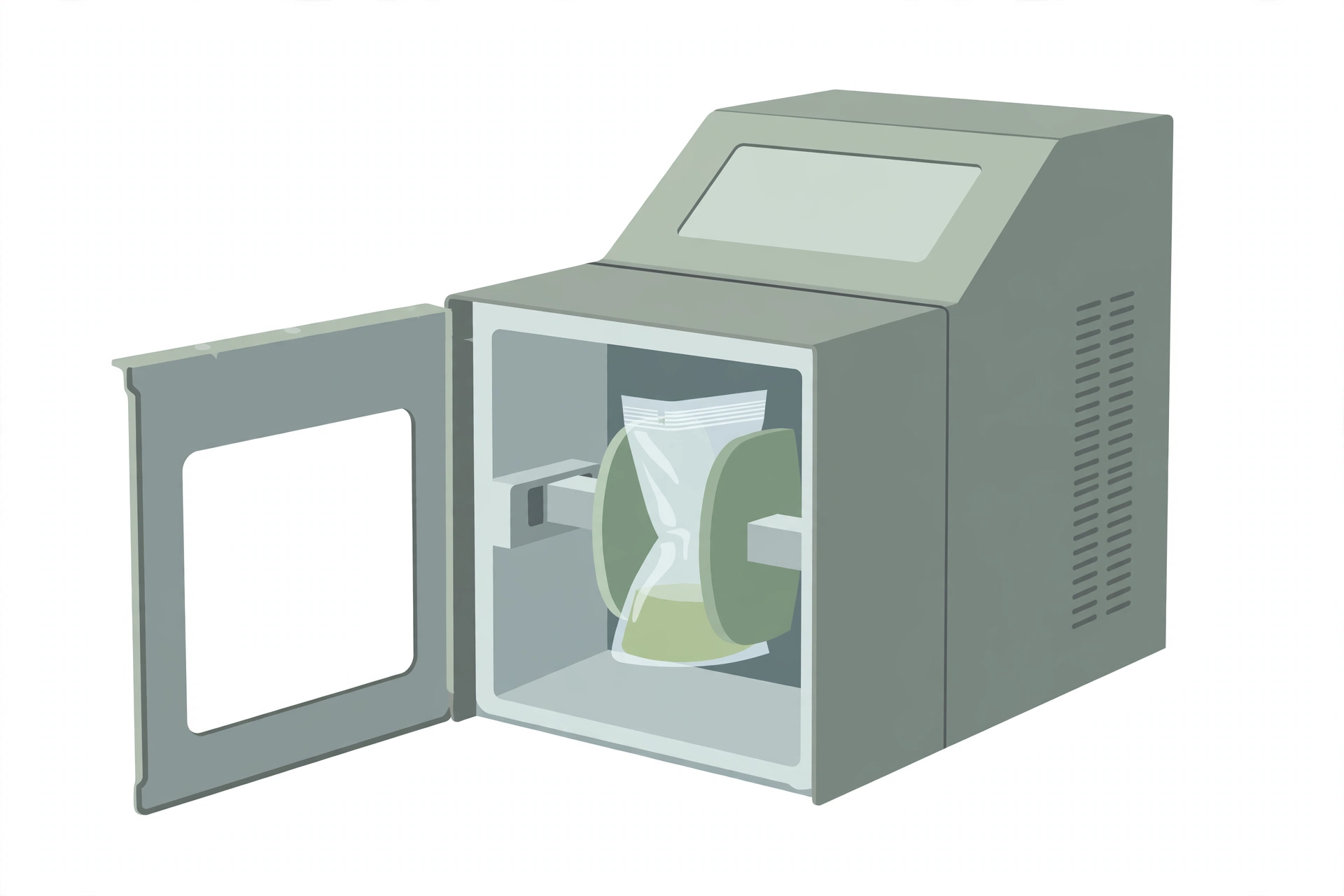
- Stomacher, blender or equivalent
- Incubators and/or waterbaths capable of maintaining the required temperatures
Reagents
- Sorbitol Bile Salts (SB) broth OR Peptone Sorbitol Bile Salts (PSB) broth (PSB broth is recommended only for samples of water or other products lacking a source of nitrogen)
- Neutralizing buffer and agar (see the Environmental Sampling method, MFLP-41)
- Phosphate Buffered Saline (see the Environmental Sampling method, MFLP-41)
- MacConkey agar
- Trypicase Soy Agar (TSA) plates and tubes
- Trypicase Soy (TSB) Broth
- Yeast Extract Rose Bengal (YER) broth
- Bile Oxalate Sorbose (BOS) broth
- Xylose Lysine Desoxycholate (XLD) agar
- Cefsulodin Irgasan Novobiocin (CIN, YSA) agar
- Kligler Iron Agar (KIA) or Triple Sugar Iron (TSI) slants
- Lysine Iron Agar (LIA) or Lysine Arginine Iron agar (LAIA) slants
- Primary screening tests: Oxidase reagent or commercial oxidase strips, Christensen’s Urea agar (CUA), Phenylalanine agar and reagent, Lysine Decarboxylase broth, Motility Test Medium
- Tube biochemical media for the differentiation of Yersinia spp.: Indole and reagent, Voges-Proskauer medium and reagents, Simmon’s Citrate, Mucate, L-ornithine, Pyrazinamidase and reagent, Sucrose, Cellobiose, L-rhamnose, Melibiose, L-sorbose, L-fucose
- Tube biochemical media for the differentiation of Y. entercolitica biotypes: Lipase, Esculin, Indole and reagent, Xylose, Salicin, Trehalose, Pyrazinamidase and reagent, β-D-glucosidase, Voges-Proskauer medium and reagent
- Magnesium-Oxalate agar
- Lipase (Tween-esterase) agar
- Mineral oil
- Alkali solution (0.5% KOH in 0.5% NaCl)
- Control cultures (ATCC or equivalent)
- Gram stain solutions
The media are commercially available and are to be prepared and sterilized according to the manufacturer’s instructions.
Media formulations
- Sorbitol Bile Salts (SB) Broth
- Basal Medium
- Sodium phosphate, dibasic, anhydrous (Na₂HPO₄) — 8.23 g
- Sodium phosphate, monobasic, monohydrate (NaH₂PO₄·H₂O) — 1.2 g
- Sodium chloride (NaCl) — 5.0 g
- Bile salts (No. 3 Difco) — 1.5 g
- D-Sorbitol — 10.0 g
- Distilled water — 1.0 L
- Complete Medium: suspend the basal medium ingredients in 1.0 L of distilled water; heat to boiling to dissolve completely; autoclave at 121 °C for 15 min
- MacConkey Agar
- Basal Medium
- Peptone — 20.0 g
- Lactose — 10.0 g
- Bile salts No. 3 — 1.5 g
- Sodium chloride (NaCl) — 5.0 g
- Neutral red — 0.03 g
- Crystal violet — 0.001 g
- Agar — 13.5 g
- Distilled water — 1.0 L
- Final pH: 7.1 ± 0.2
- Complete Medium: suspend the ingredients in 1.0 L of distilled water and heat to boiling with frequent swirling; autoclave at 121 °C for 15 min and cool to 45–50 °C; dispense the medium into plates and allow to dry with the lids partially removed
- Xylose-Lysine-Deoxycholate (XLD) Agar
- Basal Medium
- Lactose — 7.5 g
- Sucrose — 7.5 g
- Sodium thiosulfate (Na₂S₂O₃) — 6.8 g
- L-Lysine — 5.0 g
- Sodium chloride (NaCl) — 5.0 g
- Xylose — 3.75 g
- Yeast extract — 3.0 g
- Sodium deoxycholate (C₂₄H₃₉O₄Na) — 2.5 g
- Ferric ammonium citrate — 0.8 g
- Phenol red — 0.08 g
- Agar — 15.0 g
- Distilled water — 1.0 L
- pH 7.4 ± 0.2
- Complete Medium: suspend the basal medium ingredients in 1.0 L of distilled water; mix thoroughly and gently heat with agitation just until the medium boils; do not overheat — overheating causes precipitation; DO NOT AUTOCLAVE; cool to 45–50 °C and dispense into sterile Petri dishes
- Kligler Iron Agar (KIA) slants
- Basal Medium
- Pancreatic digest of casein — 10.0 g
- Peptic digest of animal tissue — 10.0 g
- Lactose — 20.0 g
- Glucose — 1.0 g
- Sodium chloride (NaCl) — 5.0 g
- Ferric ammonium citrate — 0.5 g
- Sodium thiosulfate (Na₂S₂O₃) — 0.5 g
- Agar — 15.0 g
- Phenol red — 0.025 g
- Distilled water — 1.0 L
- Final pH: 7.4 ± 0.2
- Note: when used for halophilic Vibrio species, add sodium chloride to a final concentration of 2–3%
- Complete Medium: mix all ingredients thoroughly; heat with agitation to dissolve; dispense into 13 x 100 screw-cap tubes; autoclave at 121 °C for 15 min; cool, then slant the tubes so the agar forms deep butts
- Lysine Iron Agar (LIA) slants
- Basal Medium
- Peptone — 5.0 g
- Yeast extract — 3.0 g
- Dextrose — 1.0 g
- L-Lysine — 10.0 g
- Ferric ammonium citrate — 0.5 g
- Sodium thiosulfate (Na₂S₂O₃) — 0.04 g
- Bromcresol purple — 0.02 g
- Agar — 15.0 g
- Distilled water — 1.0 L
- Final pH: 6.7 ± 0.2
- Complete Medium: suspend the ingredients in distilled water and heat to boiling with frequent swirling; dispense the appropriate volume of medium into tubes; autoclave at 121 °C for 15 min; cool in a slanted position to form deep butts
- Motility Test Medium
- Basal Medium
- Beef extract — 3.0 g
- Pancreatic digest of gelatin — 10.0 g
- NaCl — 5.0 g
- Agar — 4.0 g
- Distilled water — 1.0 L
- pH 7.2 ± 0.2
- Complete Medium: suspend the ingredients in water and mix thoroughly; heat with frequent agitation and boil for 1 min to dissolve completely; dispense and sterilize at 121 °C for 15 min
- Alkali solution (0.5% KOH in 0.5% NaCl)
- To prepare: dissolve 0.5 g of potassium hydroxide and 0.5 g of sodium chloride in distilled water and make up to 100 mL
Sample preparation
Handling of sample units
- In the laboratory prior to analysis, except for shelf-stable foods, keep sample units refrigerated or frozen depending on the nature of the product; thaw frozen samples in the refrigerator, or under time and temperature conditions which prevent microbial growth or death
- Analyses should be started as soon as possible after receipt of samples in the laboratory
- Each sample unit may be analysed individually or the analytical units taken from each sample unit may be combined as appropriate
- For environmental samples, refer to the sampling procedures given in MFLP-41
Procedure
One-step enrichment
Aseptically weigh a 25 g sample into a tared blender jar or stomacher bag and add 225 mL Sorbitol Bile Salts (SB) Broth or Peptone Sorbitol Bile Salts (PSB) Broth (or sufficient to obtain a 1:10 dilution); if analytical units other than 25 g are required, maintain a ratio of 1 part sample material to 9 parts SB or PSB; to reduce the workload, up to 5 analytical units may be composited into a single test sample (e.g. 125 g or mL)
↓
Blend for 30 seconds or stomach for 2 minutes
↓
For smaller environmental swabs (e.g., cotton tip) in transport media, transfer swab and contents into 10 mL of PSB or composite up to 5 swabs with 10 mL PSB per swab; add 100 mL of PSB per sponge or composite up to 5 sponges with 100 mL PSB per sponge; agitate thoroughly
↓
Direct plating (optional): streak 2–3 loopfuls of the homogenate onto MacConkey and Cefsulodin Irgasan Novobiocin (CIN) agar plates before incubation of the enrichment broth; incubate the MacConkey plates at 22–25 °C for 48 h and the CIN at 30 °C for 20 h; proceed to primary screening and identification if required
↓
Incubate the enrichment broths, without shaking, at 4 °C for 14 to 21 days or at 22–25 °C for 3 to 5 days, or 2 to 3 days with shaking at 100 cycles/min
Note: The PSB Broth is recommended only for samples of water or other products lacking a source of nitrogen.
Two-step enrichment (optional)
Follow the procedure described in the One-Step Enrichment, except use Yeast Extract Rose Bengal (YER) broth as the primary enrichment broth, instead of PSB or SB broths
↓
Incubate YER broth at 10 °C for 3 days (or at 4 °C for 9 days)
↓
Transfer the YER broth culture into Bile Oxalate Sorbose (BOS) broth, the secondary enrichment broth; inoculum size should be in a 1:100 ratio (generally 0.5 mL of YER broth culture to 50 mL of BOS broth)
↓
Incubate BOS broth at 22–25 °C for 5 days
Alkali treatment and primary plating
Add 0.5 mL of the enrichment culture into 4.5 mL of alkali solution (0.5% KOH in 0.5% NaCl)
↓
Shake or vortex the alkali-broth suspension and immediately (5 to 10 sec.) streak one loopful onto MacConkey and CIN agar; if an additional plating agar is desired, XLD agar may be used
↓
Incubate MacConkey and XLD plates at 22–25 °C for 36–48 h and CIN agar at 30 °C for 24 h
↓
Inoculate several known cultures onto the above plating media for controls
Primary screening
Examine plates, including controls, for colonies suggestive of Yersinia enterocolitica:
– MacConkey — 1–2 mm pale or colourless transparent colonies (lactose negative)
– XLD — 1–2 mm yellow colonies (sucrose positive, lysine decarboxylase negative, H₂S negative)
– CIN — 1–2 mm colonies with dark red centres surrounded by a transparent border
– A magnifying glass with obliquely transmitted light may be used
↓
Re-streak suspect colonies onto a fresh CIN plate to check purity and confirm typical, distinctive colonial morphology; streak a TSA plate using the same colony to obtain a suitable colony for additional tests, such as oxidase or the Vitek identification system
↓
Oxidase test: place a 2 cm square piece of filter paper in an empty Petri dish and add one drop of oxidase reagent to its centre; using a platinum loop, glass rod or wooden applicator stick, smear cells onto the reagent-impregnated paper in a spot 3–5 mm in diameter; the oxidase test is positive if the cell mass turns dark blue or purple in 30 seconds; no colour change is indicative of a negative reaction; Yersinia spp. are oxidase negative; oxidase strips are also commercially available
↓
Proceed with the following tests for oxidase negative colonies only
↓
Pick at least 2 suspect colonies from each plate and inoculate TSI or KIA agar and LAIA or LIA agar; inoculate the first tube by stabbing the butt and streaking the slant with a straight needle; repeat this procedure with the second tube without returning the needle to the original colony; incubate slants at 22–25 °C and examine after 24 h and 48 h; Yersinia spp. give the following reactions: TSI — acid slant/acid butt (A/A), gas negative, H₂S negative; KIA — alkaline slant/acid butt (K/A), gas negative, H₂S negative; LIA or LAIA — alkaline slant/acid butt (K/A), gas negative, H₂S negative
↓
Urease test: inoculate the culture to a Christensen’s Urea agar in a tube or plate by making radial streaks of each culture onto the upper part of the slant or onto one quarter to on sixth of each plate; incubate 24 h at 22–25 °C; Y. enterocolitica is urease positive (dark pink colouration); no colour change is indicative of a negative test
↓
Phenylalanine deaminase test: inoculate the oxidase-negative culture, using a loop, into a tube or plate of phenylalanine agar as described in the Urease test; after incubation at 22–25 °C for 24 h, add several drops of 10% ferric chloride solution to the growth; a positive reaction is indicated by the development of a dark green colour in the fluid and on the growth surface; no colour change is a negative test; Yersinia spp. are negative for phenylalanine deaminase
↓
Motility test: inoculate cultures, giving typical reactions with the above tests, into two tubes of motility test medium by using an inoculating needle and making one gentle stab (2 cm deep.); incubate one tube at 22–25 °C and the other at 35 °C for 48 h; most Yersinia spp. (except Y. pestis) are motile at 22–25 °C but non-motile at 35 °C
↓
Refer to Table 1 for the differential characteristics of Yersinia and related genera
Confirmatory biochemical tests
(a) Inoculate the presumptive Yersinia cultures and controls into the media listed in Table 2 and carry out the tests as described in the table
↓
(b) Differentiate the various Yersinia spp. by referring to Table 3
↓
Rapid identification kits, such as the Vitek or API 20E, or equivalent may be used for the confirmation of Yersinia spp. to the genus level; follow manufacturer’s instructions for use, including incubation temperature; these test results may be used in place of the corresponding tests in Primary screening
Serotyping (optional)
- If serotyping of Yersinia spp. is needed, facilities may be available for typing human and non-human isolates at the National Reference Centre for Yersinia, Provincial Public Health Laboratory, Public Health Ontario (the source gives the laboratory’s address and telephone number)
Biotyping (optional)
If biotyping of Y. enterocolitica is required, inoculate the media described in Table 4 (with the exception of test 8) from a fresh TSA culture which had been incubated at 22–25 °C for 24–48 h; if indole, pyrazinamidase, and Voges-Proskauer were done previously, they need not be repeated
↓
Incubate all tubes except for β-D-glucosidase, and carry out the tests as described in Table 4
↓
For β-D-glucosidase, carry out the test using a McFarland turbidity standard 3
↓
Identify the biotypes of Y. enterocolitica by referring to Table 5
In vitro pathogenicity testing (optional)
Most Y. enterocolitica isolated from environmental sources are non-pathogenic strains; only potentially virulent forms of the organism should be reported
Screening of presumably pathogenic strains
Present knowledge indicates that virulent forms are confined to some strains of Y. enterocolitica that give negative results with three tests (salicin, esculin, and pyrazinamidase)
↓
Prepare a fresh culture on TSA and incubate at 22–25 °C for 24–48 h
↓
Carry out the tests as described below:
| Test | Inoculation | Incubation °C | Incubation hours | Positive | Negative |
|---|---|---|---|---|---|
| Esculin | inoculate by stabbing | 30 | 24 | Black | No change |
| Salicin | 30 | 24 | Yellow | No change | |
| Pyrazinamidase | streak slant | 30 | 48 | Brownish-Pink | No change |
Note: These tests may be omitted if they have already been done for biotyping of Yersinia spp.
↓
If Y. enterocolitica showed negative results with esculin, salicin, and pyrazinamidase, the culture should be considered as potentially pathogenic
Confirmatory virulence tests for presumptive pathogen
Calcium Dependency Test: prepare a fresh cell suspension of approximately 10² cfu/mL in 0.85% saline; add the cell suspension to duplicate plates of both Magnesium Oxalate agar and TSA control plates; spread the inoculum over the entire surface of the media; incubate one plate of each media type at 22–25 °C and the second set of plates at 35 °C for 48 h; the reaction is regarded as positive if at 25 °C the colonies are of large and uniform size on both media but at 35 °C colonies on Magnesium Oxalate agar are a mixture of large colonies (0.5 mm to 1 mm in diameter) and small colonies (0.1 mm); the number of small colonies should be more than 20%; the cultures showing positive results are calcium dependent and are considered as presumptive pathogens or harbouring the plasmids carrying virulent factors
↓
Autoagglutination Test: prepare a fresh culture on TSA and incubate at 22–25 °C for 24–48 h; inoculate cell mass, using a loop, into a pair of tubes containing 2–4 mL of MR-VP broth (instead of the tissue culture medium with calf serum suggested by the original method); incubate 1 tube at 22–25 °C and the other at 35 °C for 24 h; a positive result consists of a confluent growth at 22–25 °C but at 35 °C there is agglutination (clumping) along the walls and/or bottom of the tube with clear fluid at the top of the tube; any other pattern for both temperatures is considered a negative test
Table 1. Differential Characteristics of Yersinia and related genera
| Characteristic | Yersinia | Hafnia | Serratia | Citrobacter | Enterobacter | Escherichia | Klebsiella | Proteus |
|---|---|---|---|---|---|---|---|---|
| Urease | + | – | d* | d | d | – | d | + |
| Motility at 22–25 °C | + | + | + | + | + | + | – | + |
| Motility at 35 °C | – | + | + | + | + | d | – | + |
| Arginine dihydrolase | – | – | – | + | d | – | – | – |
| Lysine decarboxylase | – | + | d | – | d | + | d | – |
| Phenylalanine deaminase | – | – | – | – | – | – | – | + |
| H₂S production | – | – | + | d | – | – | – | d |
* different reactions in different species
Table 2. Tube Biochemical Tests for Differentiation of Yersinia spp.
| Test | °C | Hours | Positive reaction | Negative reaction |
|---|---|---|---|---|
| Indole | 30 | 24–48 | red | no change |
| V-P | 30 | 24–48 | red | no change |
| Citrate (Simmons) | 30 | 24–48 | blue | no change |
| Mucate | 30 | 24–48 | yellow | no change |
| L-Ornithine | 30 | 24–48 | purple (violet) | yellow |
| Pyrazinamidase | 30 | 24–48 | brownish-pink | yellow |
| Sucrose | 30 | 24–48 | yellow | no change |
| Cellobiose | 30 | 24–48 | yellow | no change |
| L-Rhamnose | 30 | 24–48 | yellow | no change |
| Melibiose | 30 | 24–48 | yellow | no change |
| L-Sorbose | 30 | 24–48 | yellow | no change |
| L-Fucose | 30 | 24–48 | yellow | no change |
Note: Read all carbohydrate utilization tests at 24 and 48 hours.
Note: After inoculation of L-ornithine, overlay with 1–2 mL of sterile mineral oil. This test requires an uninoculated control broth.
Table 3. Biochemical Differentiation of Yersinia species
| Test | Y. enterocolitica | Y. intermedia | Y. frederiksenii | Y. kristensenii | Y. aldovae | Y. rhodei | Y. mollaretti | Y. bercovieri | Y. pseudotuberculosis |
|---|---|---|---|---|---|---|---|---|---|
| Indole | d^a | + | + | d | – | – | – | – | – |
| Voges-Proskauer | d | + | d | – | + | – | – | – | – |
| Citrate (Simmons) | – | + | d | – | d | + | – | – | – |
| L-Ornithine | + | + | + | + | + | + | + | + | – |
| Mucate, acid | – | d | d | – | d | – | + | + | – |
| Pyrazinamidase | d | + | + | + | + | + | + | + | – |
| Sucrose | + | + | + | – | – | + | + | + | – |
| Cellobiose | + | + | + | + | – | + | + | + | – |
| L-Rhamnose | – | + | + | – | + | – | – | – | + |
| Melibiose | – | + | – | – | – | d | – | – | + |
| L-Sorbose | d | + | + | + | – | ND^b | + | – | – |
| L-Fucose | d | d | + | d | d | ND | – | + | – |
a Different reactions;
b Not determined.
Table 4. Biochemical Tests for Differentiation of Biotypes of Y. enterocolitica
| Test | °C | Hours | Positive | Negative |
|---|---|---|---|---|
| (1) Lipase | 30 | 24–48 | clear zone | no clear zone |
| (2) Esculin | 30 | 24–48 | black | no change |
| (3) Indole | 30 | 24–48 | red | no change |
| (4) Xylose | 30 | 24–48 | yellow | no change |
| (5) Salicin | 30 | 24–48 | yellow | no change |
| (6) Trehalose | 30 | 24–48 | yellow | no change |
| (7) Pyrazinamidase | 30 | 24–48 | brownish-pink | no change |
| (8) β-D-glucosidase | 30 | 24 | yellow | no change |
| (9) Voges-Proskauer | 30 | 24–48 | red | no change |
Table 5. Biotyping Scheme for Y. enterocolitica
| Biochemical Test | 1A | 1B | 2 | 3 | 4 | 5 | 6^b |
|---|---|---|---|---|---|---|---|
| Lipase (Tween-esterase) | + | + | – | – | – | – | – |
| Hydrolysis of esculin | +/- | – | – | – | – | – | – |
| Indole | + | + | (+) | – | – | – | – |
| Acid from xylose | + | + | + | + | – | V | + |
| Acid from salicin | +/- | – | – | – | – | – | – |
| Acid from trehalose | + | + | + | + | + | – | + |
| Pyrazinamidase | + | – | – | – | – | – | + |
| β-D-glucosidase | + | – | – | – | – | – | – |
| Voges-Proskauer | + | + | + | +/-^a | + | (+) | – |
a A biotype of serotype O:3 found in Japan.
b Recently recognized as two new species, Y. mollaretti and Y. bercovieri
+ = positive reaction; − = negative reaction; V = variable reaction; ( ) = delayed reaction.
Precautions
- The laboratory Supervisor must ensure that completion of the analysis described in this method is done in accordance with the International Standards reference “ISO/IEC 17025:1999 (or latest version): General Requirements for the Competence of Testing and Calibration Laboratories”
- If the analyst uses any variations of the media listed here (either product that is commercially available or made from scratch), it is the responsibility of the analyst or Laboratory Supervisor to ensure equivalency
- It is the responsibility of each laboratory to ensure that the temperatures of the incubators or water baths are maintained at the recommended temperatures; where 35 °C is recommended in text of the method the incubator may be at 35 ± 1.0 °C; similarly, lower temperatures of 30 or 25 may be ± 1.0 °C; however, where higher temperatures are recommended, such as 43 or 45.5 °C, it is imperative that the incubators or water baths be maintained within 0.5 °C due to potential lethality of the higher temperatures on the microorganism(s) being isolated
References
- Health Canada, Health Products and Food Branch. Isolation of Yersinia enterocolitica from Foods and Environmental Samples. Compendium of Analytical Methods, Volume 3, Laboratory Procedure MFLP-48, May 2012.
- Atlas, R.M. 1997. Handbook of Microbiological Media. Second edition. L.C. Parks (editor). CRC Press Inc.
- Bergey’s Manual of Determinative Bacteriology. 1994. Ninth edition. J.G. Holt, N.R. Krieg, P.H.A. Sneath, J.T. Staley and S.T. Williams (eds), Williams and Wilkins, Baltimore,MD.
- Gemski, P., J.R. Lazere and T. Casey. 1980. Plasmid associated with pathogenicity and calcium dependency of Yersinia enterocolitica. Infect. Immun. 27:682.
- Laird, W.J. and D.C. Cavanaugh. 1980. Correlation of autoagglutination and virulence of Yersiniae. J. Clin. Microbiol. 11:430.