SOP: Isolation and Identification of Shigella spp. from Foods
GIFSQ — Global Institute for Food Safety & Quality
Principle
A portion of the food sample is enriched in a selective broth containing novobiocin, then plated onto selective-differential agars for the isolation of Shigella spp. Presumptive Shigella isolates are screened and identified by biochemical testing and serology. The method is based on the FDA Bacteriological Analytical Method for Shigella, with some modifications. Screening and/or confirmation by the PCR method (MFLP-26) is optional.
This method applies to the detection of viable Shigella spp. from fresh produce and commercial salads, including spinach, parsley, Greek salad, four bean salad, Greek pasta salad, Romaine lettuce, fresh mint, green onion, celery, softshell clams and strawberries. It can be used to determine compliance with Sections 4 and 7 of the Food and Drug Act. This revised method replaces MFLP-25, dated February 2004.
Apparatus
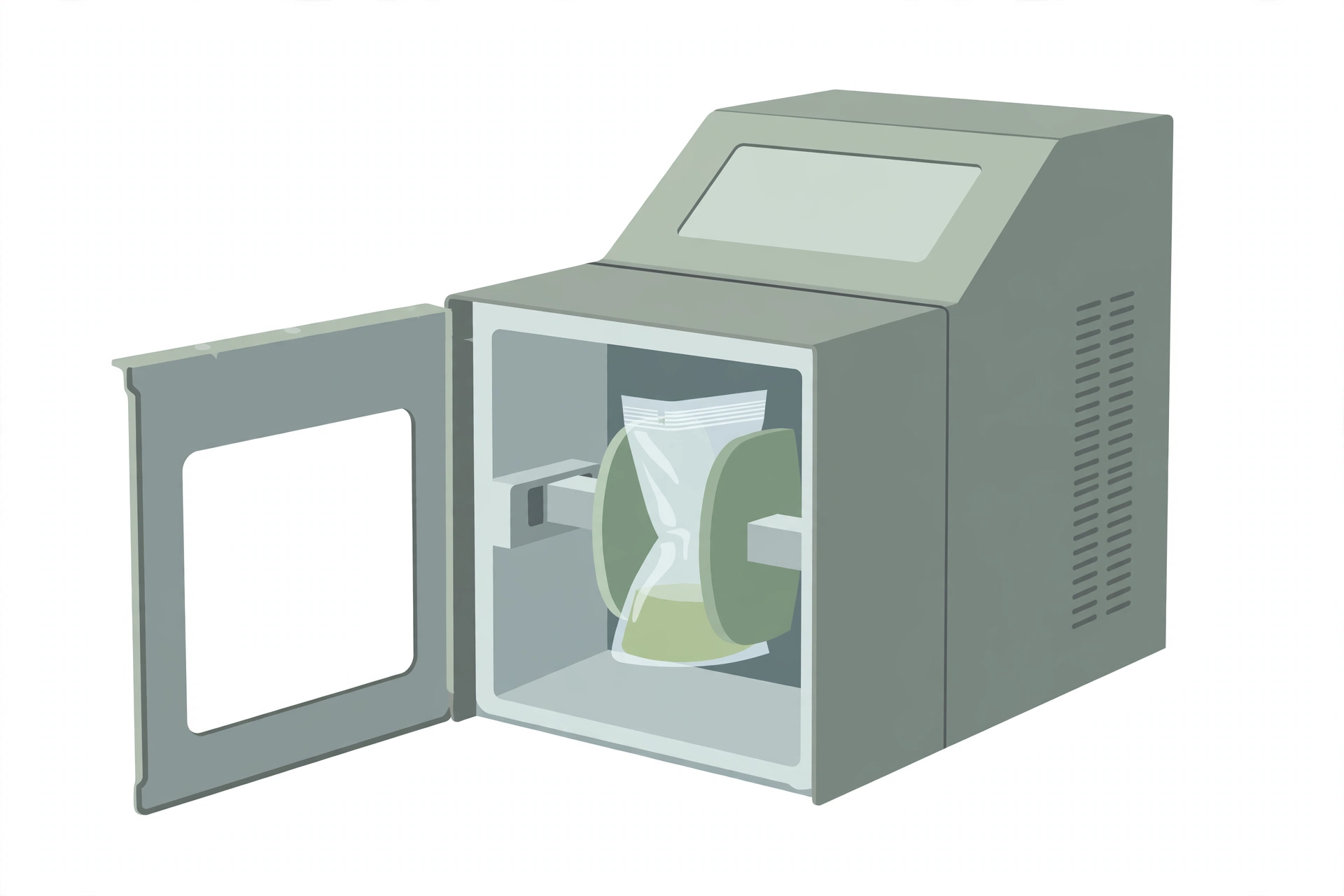
- Stomacher filter bags
- Stomacher bags
- Erlenmeyer flasks (optional — instead of stomacher bags for the enrichment step)
- pH meter or paper capable of distinguishing 0.2 to 0.3 pH units within a range of 6.0 to 8.0 (available from EM Science or J.T. Baker through VWR)
- CO₂ incubator, or anaerobic jars with CO₂ generator bags
- Incubators capable of maintaining 35 °C and 42 °C
Reagents
The media below are commercially available and are prepared and sterilized according to the manufacturer’s instructions.
- Shigella broth
- To prepare: basal medium — 20.0 g tryptone, 2.0 g potassium phosphate, dibasic (K₂HPO₄), 2.0 g potassium phosphate, monobasic (KH₂PO₄), 5.0 g sodium chloride (NaCl), 1.0 g glucose and 1.5 mL Tween 80 per 1.0 L of distilled water; pH 7.0 ± 0.2. Suspend the basal ingredients in 1.0 L of distilled water, heat to boiling to dissolve completely, and autoclave at 121 °C for 15 min. Cool to 50 °C, then aseptically add 2.5 mL of the novobiocin solution to 225 mL of basal medium and mix thoroughly. Novobiocin solution: suspend 50 mg of novobiocin in 1.0 L of distilled water and sterilize by filtration through a 0.45 µm membrane filter.
- MacConkey agar (MAC)
- To prepare: suspend 20.0 g peptone, 10.0 g lactose, 1.5 g bile salts No. 3, 5.0 g sodium chloride (NaCl), 0.03 g neutral red, 0.001 g crystal violet and 13.5 g agar in 1.0 L of distilled water and heat to boiling with frequent swirling; final pH 7.1 ± 0.2. Autoclave at 121 °C for 15 min, cool to 45–50 °C, dispense into plates and allow to dry with the lids partially removed.
- Xylose lysine desoxycholate (XLD) agar
- To prepare: suspend 7.5 g lactose, 7.5 g sucrose, 6.8 g sodium thiosulfate (Na₂S₂O₃), 5.0 g L-lysine, 5.0 g sodium chloride (NaCl), 3.75 g xylose, 3.0 g yeast extract, 2.5 g sodium deoxycholate, 0.8 g ferric ammonium citrate, 0.08 g phenol red and 15.0 g agar in 1.0 L of distilled water; pH 7.4 ± 0.2. Mix thoroughly and gently heat with agitation just until the medium boils — do not overheat (overheating causes precipitation). DO NOT AUTOCLAVE. Cool to 45–50 °C and dispense into sterile Petri dishes.
- Hektoen agar (HE)
- To prepare: suspend 12.0 g lactose, 12.0 g peptic digest of animal tissue, 12.0 g sucrose, 9.0 g bile salts, 5.0 g sodium chloride (NaCl), 5.0 g sodium thiosulphate (Na₂S₂O₃), 3.0 g yeast extract, 2.0 g salicin, 1.5 g ferric ammonium citrate, 0.1 g acid fuchsin, 0.064 g bromthymol blue and 13.5 g agar in 1.0 L of distilled water; pH 7.6 ± 0.2. Heat to boiling with constant stirring to dissolve; do not autoclave; dispense into sterile Petri dishes. Caution: acid fuchsin is a potential carcinogen — avoid inhaling the powdered dye and avoid contact with skin; wear suitable protective clothing.
- Triple sugar iron Agar (TSI)
- To prepare: suspend 3.0 g beef extract, 3.0 g yeast extract, 15.0 g peptone, 5.0 g proteose peptone, 10.0 g lactose, 10.0 g sucrose, 1.0 g dextrose, 0.2 g ferrous sulfate (FeSO₄·7H₂O), 5.0 g sodium chloride (NaCl), 0.3 g sodium thiosulfate (Na₂S₂O₃), 12.0 g agar and 0.024 g phenol red in 1.0 L of distilled water; pH 7.4 ± 0.2. Heat to boiling to dissolve completely, dispense 8 mL into tubes, autoclave at 121 °C for 15 min, and cool the tubes in a slanted position to form deep butts.
- Lysine iron agar (LIA)
- To prepare: suspend 5.0 g peptone, 3.0 g yeast extract, 1.0 g dextrose, 10.0 g L-lysine, 0.5 g ferric ammonium citrate, 0.04 g sodium thiosulfate (Na₂S₂O₃), 0.02 g bromcresol purple and 15.0 g agar in distilled water and heat to boiling with frequent swirling; final pH 6.7 ± 0.2. Dispense the appropriate volume of medium into tubes, autoclave at 121 °C for 15 min, and cool in a slanted position to form deep butts.
- Motility test medium with 2,3,5-triphenyl tetrazolium chloride (TTC)
- To prepare the basal medium: suspend 3.0 g beef extract, 10.0 g pancreatic digest of gelatin, 5.0 g NaCl and 4.0 g agar in 1.0 L of distilled water (pH 7.2 ± 0.2); heat with frequent agitation and boil for 1 min to dissolve completely; dispense and sterilize at 121 °C for 15 min.
- Trypticase soy agar (TSA) or equivalent
- To prepare: suspend 15.0 g tryptone, 5.0 g soya peptone, 5.0 g sodium chloride (NaCl) and 15.0 g agar in 1.0 L of distilled water (pH 7.3 ± 0.2); heat to boiling with constant stirring to dissolve, without overheating; autoclave at 121 °C for 15 min.
- Trypticase soy broth (TSB) or equivalent
- To prepare: suspend 17.0 g of tryptone, 3.0 g of soya peptone, 2.5 g of dextrose, 5.0 g of sodium chloride (NaCl) and 2.5 g of dipotassium phosphate (K₂HPO₄) in 1.0 L of distilled water (pH 7.3 ± 0.2); heat to boiling with constant stirring to dissolve, dispense, and autoclave at 121 °C for 15 min.
- Physiological saline, 0.85% (sterile)
- To prepare: dissolve 8.5 g of sodium chloride (NaCl) in distilled water and make up to 1.0 L; sterilize at 121 °C for 15 min.
- Novobiocin — a sterile novobiocin solution is prepared so the Shigella broth contains 0.5 µg of novobiocin per mL
- Novobiocin solution: suspend 50 mg of novobiocin in 1.0 L of distilled water and sterilize by filtration through a 0.45 µm membrane filter; aseptically add 2.5 mL to 225 mL of cooled Shigella broth basal medium (see the Shigella broth preparation above).
- Sterile 1 N HCl and sterile 1 N NaOH
- To prepare sterile 1 N HCl: add 83 mL of concentrated hydrochloric acid to distilled water and make up to 1.0 L; sterilize. To prepare sterile 1 N NaOH: dissolve 40 g of sodium hydroxide in distilled water and make up to 1.0 L; sterilize.
- Identification test kits (API, VITEK, Micro-ID) or equivalent
- Polyvalent Shigella antisera for groups A, A1, B, C, C1, C2 and D (or send isolates to a Reference Laboratory Typing Centre)
Sample preparation
- Analyse samples as soon as possible, preferably within 24 h of receipt. During transport, with the exception of shelf-stable products, keep the sample units refrigerated or frozen depending on the nature of the product. Thaw frozen samples in a refrigerator, or under time and temperature conditions which prevent microbial growth or death. Foods which are partially thawed are stored in a refrigerator.
- Use appropriate containers to ensure that drippings from the product do not contaminate the laboratory environment.
- If the sample unit received for analysis is less than the recommended analytical unit, analyse the entire amount and record the weight used. Adjust the volume of enrichment broth to maintain a 1 in 10 dilution of the sample.
- Use aseptic techniques and sterile equipment at all stages of analysis. Containment during the handling of powdered products is critical if cross-contamination of the work environment is to be avoided.
- Have ready sterile Shigella broth and sterile novobiocin solution.
- Clean the surface of the working area with a suitable disinfectant.
- Prepare and verify the suitability of all controls. Include positive and reagent controls. Shigella sonnei (ATCC 29930), Shigella flexneri (ATCC 29903), Shigella dysenteriae (ATCC 13313) or Shigella boydii (ATCC 8700) are included as a positive control. (ATCC numbers are included as reference; equivalent Shigella strains may also be used.)
- To ensure a truly representative analytical unit, agitate liquids or free flowing materials until the contents are homogeneous. If the sample unit is a solid, obtain the analytical unit by taking a portion from several locations within the sample unit. To reduce the workload the analytical units may be combined for analysis; it is recommended that a composite contain no more than five analytical units.
Procedure
Enrichment of Shigella spp.
- Aseptically weigh 25 g or mL (the analytical unit) into a filter stomacher bag and add 225 mL of Shigella broth containing 0.5 µg of novobiocin per mL
↓ - Hold the suspension 10 min at room temperature and periodically shake or gently massage the bag
↓ - Pour the supernatant into a sterile 500 mL Erlenmeyer flask or into a new stomacher bag
↓ - Adjust the pH of the supernatant, if necessary, to 7.0 ± 0.2 with sterile 1 N NaOH or 1 N HCl
↓ - Place the flasks or stomacher bags in a CO₂ incubator or into anaerobic jars containing CO₂ generator bags; incubate at 42 °C for 20 h ± 2 h
↓ - Set up a positive and a negative cultural control at the time of food sample analysis
↓ - After incubation, screen the enrichment broth for Shigella spp. by the PCR technique (optional — see MFLP-26) and/or proceed with selective isolation
↓ - If the PCR method is being used in lieu of cultural screening, refrigerate the enrichment broth; if the PCR is positive for Shigella spp., proceed to selective isolation using the refrigerated broth
(When analysing larger volumes, the enrichment broth is pre-warmed to 35 °C.)
Selective isolation
- Agitate the enriched broth and streak replicate loopfuls onto at least 2 selective-differential media — MacConkey (low selectivity), XLD (high selectivity) and HE (moderate to high selectivity) agars — to obtain well isolated colonies; the enriched broth may be streaked onto TSA for background control purposes only
↓ - Keep the enrichment broth refrigerated until the end of the food analysis
↓ - Incubate the plates at 35 °C for 24 ± 2 h
↓ - Examine the selective-differential plates for characteristic Shigella colonies (see the colony appearance table)
↓ - Pick suspect colonies to be confirmed by biochemical tests and/or PCR and serology
Colony appearance on selective plating media
| Organism | Selective agars | Colony appearance | Size of colonies |
|---|---|---|---|
| Shigella | MAC | Convex, colourless (lactose non-fermentor); S. sonnei colonies are flat with rough edges | 2–3 mmᵃ |
| Shigella | XLD | Red or colourless | 1–2 mmᵃ |
| Shigella | HE | Green | 2–3 mmᵃ |
| E. coli | MAC | Flat pink colonies (lactose fermentor) surrounded by a darker pink region | — |
| E. coli | XLD | Yellow | — |
| E. coli | HE | Pink to orange | — |
ᵃ S. dysenteriae 1 colonies may be smaller on MAC and on HE; and on XLD agar are frequently very tiny, unlike other Shigella species. (The source prints the sizes as “2-3 mma” and “1-2 mma”, where “a” is the footnote marker, verified against the page image.)
Screening (biochemistry and/or PCR)
- PCR: because the PCR method for Shigella is very specific, isolation of pure colonies is not necessary for this test
↓ - Biochemical screening:
- If the colonies are well isolated: select 3–5 suspicious Shigella colonies (or more if deemed necessary) per plate for biochemical screening; circle all suspicious colonies before refrigerating the plates in case additional colonies are required for testing
↓ - If the colonies are not well isolated: streak suspect colonies onto MAC for purification and incubate the plates at 35 °C for 24 ± 2 h
↓ - With a sterile needle, pick suspect colonies and inoculate into TSI, LIA and motility test medium; in parallel, test pure cultures of Shigella and E. coli strains as positive and negative controls, respectively; store the plates in the refrigerator for possible further use
↓ - Incubate the biochemical test media at 35 °C for 20–24 h, with the tubes loosely capped (erroneous biochemical results may be obtained if tubes are not loosely capped during incubation)
↓ - Interpret the reactions (see the screening reactions table): typical Shigella spp. are motility, H₂S, lysine and gas negative; however, S. dysentariae 3, S. boydii 13 and 14, and S. flexneri 6 may produce some gas
↓ - If the original suspect colonies test negative for Shigella spp. and further picking is deemed necessary — for example, when the PCR is Shigella positive and the colonies picked were biochemically negative, or for a high priority sample — additional colonies are tested
↓ - Commercially available diagnostic kits (API, VITEK, Micro-ID or equivalent) may be used to obtain detailed biochemical profiles of bacterial isolates; when using rapid diagnostic kits, follow the manufacturer’s instructions, including the temperature of incubation indicated
↓ - If the presence of Shigella is suspected, proceed with serological testing or send to a Reference Laboratory Typing Centre; if polyvalent serology has not yet been performed but the rapid method results are indicative of Shigella spp., a tentative identification can be made
↓ - If none of the isolates from a particular analytical unit are suggestive of Shigella, the analytical unit is considered to be free of Shigella
↓ - If the serological testing is not to be performed within 72 h, inoculate suspect isolates onto nutrient agar (NA) slants and incubate at 35 °C for 24 h; store the agar slants in a refrigerator; NA slants that have been stored for more than 72 are not used for serological testing — prepare fresh agar slants for this purpose
Screening reactions for Shigella spp.
| Screening medium | Shigella spp. reaction | E. coli reaction |
|---|---|---|
| TSI | K/A, no gas production (red slant/yellow butt)ᵃ | A/A, gas production (yellow slant/yellow butt with gas pockets) |
| H₂S on TSI | Negative | Negative |
| LIA | K/A (purple slant/yellow butt)ᵇ | K/K (purple slant/purple butt) |
| Motilityᶜ | Negative | Positive |
- ᵃ K = alkaline (red); A = acid (yellow); some strains of S. flexneri serotype 6, S. boydii serotypes 13 and 14 and S. dysenteriae serotype 3 produce gas from glucose.
- ᵇ K = alkaline (purple); A = acid (yellow); an alkaline reaction (purple) in the butt of the medium indicates that lysine was decarboxylated. An acid reaction (yellow) in the butt of the medium indicates that lysine was not decarboxylated.
- ᶜ Motility test medium with added TTC: positive — a diffused reddish colour throughout the medium; negative — a reddish line along the inoculation site only.
Serological identification
- Mark the following sections on an agglutination plate: C− (negative control) and T (test culture); if a Shigella control culture is available for each somatic group tested, prepare C+ (positive control) for each group
↓ - Add one drop of physiological saline to each of the areas marked T and C+, and two drops to the area marked C−
↓ - Remove sufficient culture material from a triple sugar iron, lysine iron or nutrient agar slant to prepare a heavy suspension in the test area and in the negative control area; the inoculum is withdrawn from the slope portion of the agar slants
↓ - Prepare somatic group antiserum as directed by the manufacturer; add one drop to each of the areas marked T and C+
↓ - Mix the culture-saline-antiserum suspensions in T and C+ and the saline-culture mixture in C− with a sterile needle or loop; tilt the slide preparation back and forth for 1 min
↓ - Hold the slide against a dark background and observe for agglutination; Shigella cultures usually agglutinate within 1 min
↓ - The serological test for a given culture is invalidated if the negative control shows agglutination (autoagglutination)
↓ - Isolates that react serologically and show no conflicting results in the biochemical screening tests are reported as positive for Shigella
(As some Shigella commercial antiserum is labelled or packaged differently, when using newly purchased antisera, read the package insert or check with the manufacturer if the label is not self-explanatory. If the serological test is positive, the culture is sent to a Shigella typing centre for complete serotyping. It should be stressed that any non-agglutinating culture possessing the biochemical reactions suggestive of Shigella is sent to a reference typing centre for identification. If polyvalent antisera are available and test positive but monovalent antisera for further speciation are not available, the culture is sent to a reference typing centre for complete serotyping.)
Serological differentiation of Shigella spp.
| Test | S. dysenteriae | S. flexneri | S. boydii | S. sonnei |
|---|---|---|---|---|
| Polyvalent antisera | A, A1 | B | C, C1, C2 | D |
Precautions
- The Laboratory Supervisor must ensure that the analysis described in this method is carried out in accordance with the International Standard referred to as “ISO/IEC 17025:2005 (or latest version): General Requirements for the Competence of Testing and Calibration Laboratories”.
- It is the responsibility of each laboratory to ensure that the temperature of the incubators or waterbaths is maintained at the recommended temperatures. Where 35 °C is recommended in the text of the method, the incubator may be 35 ± 1.0 °C. Similarly, lower temperatures of 30 or 25 °C may be ± 1.0 °C. However, where higher temperatures are recommended, such as 43 or 45.5 °C, it is imperative that the incubators or waterbaths be maintained within 0.5 °C due to potential lethality of higher temperatures on the microorganism being isolated.
- If the analyst uses any variations of the media listed here (either product that is commercially available or made from scratch), it is the responsibility of the analyst or Laboratory Supervisor to ensure equivalency.
- Erroneous biochemical results may be obtained if tubes are not loosely capped during incubation.
References
- Health Canada. Isolation and Identification of Shigella spp. from Foods. Compendium of Analytical Methods, Volume 3, Laboratory Procedure MFLP-25, March 2006. Research Division, Health Products and Food Branch, Health Canada.
- AOAC. 1995. Shigella. Chapter 6. In: Bacteriological Analytical Methods, 8th edition (1998), Gaitherburg, MD.
- American Public Health Association. 2001. Shigella. Chapter 38. In: Compendium of Methods for the Examination of Foods, 4th edition. F.P. Downes and K. Ito (eds.), Washington, D.C.
- Centers for Disease Control and Prevention. 1999. Isolation and identification of Shigella. Chapter 4. In: Laboratory methods for diagnosis of epidemic dysentery and cholera, Atlanta, Georgia.
- ISO/TC 34/SC9. 2002. Microbiology of food and animal feeding stuffs — Horizontal method for the detection of Shigella sp. (the source prints “ISO/DIS2 1567”)
- Kelly, A.T. and M. Fulton. 1953. Use of triphenyl tetrazolium chloride in motility test medium. Am. J. Clin. Path. 23:512.