SOP: Isolation and Identification of Anisakid Roundworm Larvae in Fish
GIFSQ — Global Institute for Food Safety & Quality
1. Principle
Roundworm larvae are separated from fish tissues by manual, sedimentary or chemical means — non-destructive candling and ultraviolet illumination, or destructive elution and digestion — and identified from their gross morphological features under the microscope, in live or preserved specimens. Candling is the procedure used in routine inspections at federally regulated processing plants. Ultraviolet illumination detects larvae at or near the flesh or viscera surface by their fluorescence; it kills live parasites, and detection is improved by a preliminary freeze/thaw of the specimen. In elution, live parasites migrate out of the tissues into saline; in digestion, all parasites — live or dead — are isolated. Digestion can recover 175% more pathogenic worms (Anisakis and Pseudoterranova) than elution, so a combination of candling, elution and digestion is recommended for best parasite yield.
2. General characteristics of anisakid roundworms
Anisakid roundworms are a group of closely related genera within the phylum Nematoda, with a relatively complex life cycle. Eggs released from mature worms pass in the faeces of marine mammals (the definitive hosts), sink to the sea floor, and hatch into second-stage larvae within days or weeks, depending on water temperature. The larvae develop to the third stage after ingestion by marine crustaceans; when a fish or squid eats the crustacean, the larvae migrate into the tissues of this second intermediate host and reach the advanced third stage on the viscera or in the muscle. When a marine mammal eats the infected fish, the larvae are released in the stomach or intestine and moult further into fourth-stage larvae and adults. Humans are accidental hosts only, with no influence on transmission.
Larvae of two anisakid roundworms — Anisakis simplex (herring worm) and Pseudoterranova decipiens (seal worm) — are implicated in the majority of human anisakiasis cases. The infective third-stage larvae occur on the viscera or in the flesh of commercially important fish species including salmon, cod, herring and mackerel, as well as squid; P. decipiens is also found in smelt and various flatfish. Two other anisakid roundworms, Contracaecum osculatum and Phocascaris spp., also occur in commercially important marine fishes and may be of public health concern.
3. Apparatus required
Candling
- Candling light table (construction described in the candling procedure)
- Small forceps
- Culture dish containing normal saline
Ultraviolet light illumination
- Hand-held or fixed ultraviolet light illuminator
- Face shield or goggles (UV light resistant)
- Small forceps
- Culture dish containing normal saline
Elution
- U.S. Standard No. 4 sieve (or a similar sized kitchen sieve)
- Large funnel (500 mL or larger) or other vessel
- Clear, flexible tubing and tubing clamp
- Ring clamp with support rod and base
- 250 mL beaker
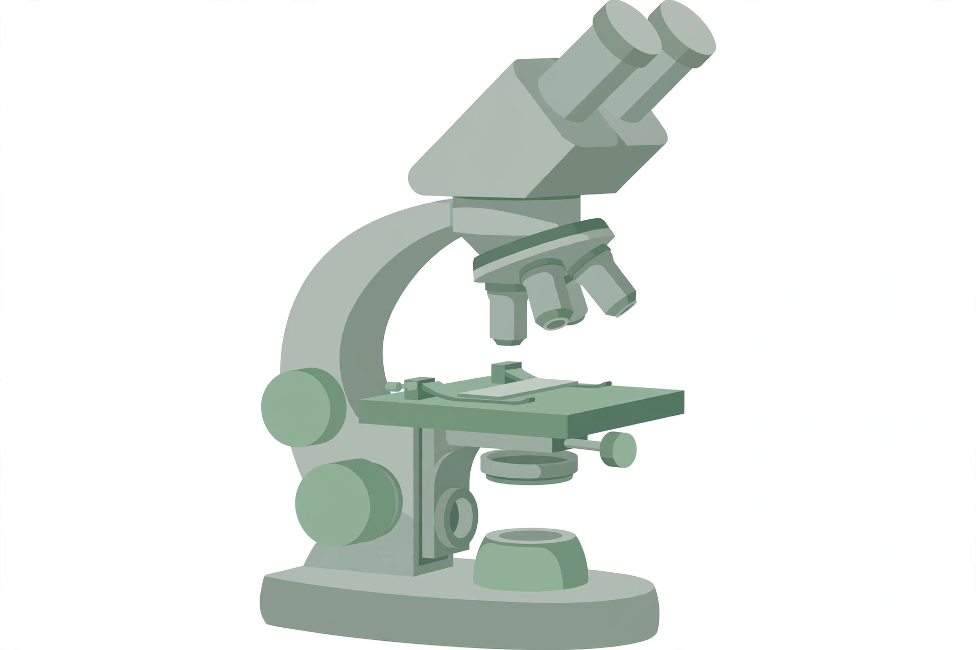
- Stereoscopic microscope or magnifier
- Forceps
- Culture dish containing normal saline
Digestion
- 1.5 L beaker; 250 mL beaker
- Shaking water bath (37°C), or bath with mechanical stirrer
- pH meter, or pH paper, and 6 N HCl
- Aluminum foil
- U.S. Standard No. 18 sieve (or a similar sized kitchen sieve)
- Large container to hold the sieve for collecting sieved material, or a U.S. Standard No. 140 sieve
- Funnel apparatus as described for elution
- Stereoscopic microscope or magnifier
- Forceps
- Culture dish containing normal saline
Preservation and storage
- Small glass vials
4. Reagents
- Normal saline: 0.85 g NaCl per 100 mL distilled water
- Pepsin solution: 15 g pepsin (1:10,000; Sigma Chemical Co.) dissolved in 750 mL normal saline
- Fixatives: glacial acetic acid, or alcohol-formalin-acetic acid (AFA) — 85 mL 85% ethanol + 10 mL formalin + 5 mL glacial acetic acid
- Preservatives: 70% ethanol, or alcohol-glycerol (9 parts 70% ethanol to 1 part glycerol)
- Clearing agents: glacial acetic acid, or glycerol, or xylene
Reagent preparations
- Glacial Acetic Acid (fixative)
- Use as supplied
- Alcohol-Formalin-Acetic Acid (AFA) fixative
- To prepare: mix 85 mL of 85% ethanol, 10 mL of formalin and 5 mL of glacial acetic acid
- 70% Ethanol (preservative)
- To prepare: mix 70 parts ethanol with 30 parts distilled water (v/v)
- Alcohol-Glycerol (preservative)
- To prepare: mix 9 parts of 70% ethanol with 1 part of glycerol
5. Sample collection
These procedures apply to the isolation and identification of live third-stage anisakid roundworm parasites in the flesh or on the viscera of a variety of commercially important marine fishes. They are intended for laboratory and inspection staff — for small quantities of fish from consumer complaints as well as larger samples from routine inspections.
6. Procedure
Candling
The recommended light table has at least two 20 watt “cool white” fluorescent tubes, held by a rigid framework below a white, translucent acrylic plastic (or other suitable material) with a translucency of 45–60%. The working surface is about 30 × 60 cm and 5–6 mm thick; the average light intensity is 1500–1800 lux measured 30 cm above the centre of the sheet, with overhead illumination of at least 500 lux (a simple photographic light table also gives adequate results)
↓
Place the fillet on the light table and examine for tightly coiled, encapsulated larvae, which appear as dark spots — unencapsulated larvae may also be seen on the fillet surface
↓
Use forceps to transfer any visible worms to a dish of normal saline
Note (per the source): candling works equally well with fresh or previously frozen fillets; its efficiency depends largely on fillet thickness; it is generally considered a costly and inefficient means of detecting parasites, and it cannot distinguish live from dead parasites.
Ultraviolet light illumination
Put on a UV-resistant face shield or safety goggles
↓
In a darkened room, hold the UV light about 10 cm above the fillet surface and examine for fluorescent worms — fluorescence colour aids preliminary identification: Anisakis and Pseudoterranova larvae fluoresce bright bluish-white, Contracaecum larvae fluoresce yellow
↓
Use forceps to transfer the worms to a dish of normal saline
Note (per the source): ultraviolet light kills any live parasites, and the procedure cannot distinguish live from dead; detection is improved by a preliminary freeze/thaw of the specimen.
Elution
Fit a U.S. Standard No. 4 sieve (or an equivalent kitchen sieve) into a large funnel or other vessel; attach tubing to the funnel neck and close it tightly with a tubing clamp
↓
Transfer up to 200 g of fish tissue to the sieve
↓
Fill the funnel or vessel with normal saline until the fish tissue is completely immersed
↓
Cover with plastic wrap or aluminum foil and leave the apparatus overnight at room temperature — live larvae migrate out of the tissues and fall through the sieve into the funnel neck (or the bottom of the vessel)
↓
After 16–18 h, drain 100 mL of sediment into a beaker
↓
With forceps, move any visible worms to a culture dish containing normal saline
↓
Carefully examine the remaining suspension with a stereoscopic microscope (120×) or magnifier for any smaller worms
Digestion
Add about 200 g of fish tissue to 750 mL of pepsin solution at 37°C in a large beaker
↓
Place the beaker in a 37°C shaking water bath so the water level is within 1 cm of the fluid level in the beaker
↓
Shake at low speed for about 15 min
↓
Adjust the sample to pH 2 with 6 N HCl, cover the beaker with foil, and continue shaking for 24 h (or until the tissue is fully digested)
↓
Pour the digested material through a U.S. Standard No. 18 sieve (or an equivalent kitchen sieve) into a suitable container
↓
Rinse the sieve with normal saline and examine the washed remains by immersing the mesh in clean normal saline
↓
Use forceps to transfer the worms to a culture dish containing normal saline
↓
Examine the material that passed through the sieve: transfer it to a clamped funnel, let it settle for 1 h, and drain the sediment into a beaker as described for elution; examine for smaller worms with a stereoscopic microscope (120×) or magnifier
Alternative — pass the digested material through nested U.S. Standard No. 18 and No. 140 sieves, separate the sieves, and immerse the screen portions in normal saline; collect the larger worms caught in the No. 18 sieve and the smaller worms caught in the No. 140 sieve, transferring each to a dish of normal saline
7. Recording results
- Record every isolated parasite as live, dead (intact), or fragments; a parasite is considered live if it shows any movement during the isolation procedures (anisakid larvae become much more active as they approach room temperature)
- Record the number of parasites per kg of product, with a preliminary identification of the parasites (or a brief description) where possible
- Record the fish species examined, its source, the total sample weight, and the weight and number of the individual samples (fillets), plus any other relevant information
8. Preservation and storage of larvae
Fix live or previously frozen worms in glacial acetic acid or AFA for 1 min, then preserve them in small glass vials containing 70% ethanol — or, preferably, an alcohol-glycerol solution. Worms may be stored in this solution indefinitely. Label each vial adequately (parasite identification, host species, location in host, sample number, date of recovery, name of examiner, and so on).
9. Parasite identification
If needed, use a clearing agent such as xylene to make the nematode cuticle more transparent. Prepare a temporary slide mount for microscopic examination, then return the worms to the preservative solution (nematodes are generally not stained for microscopic examination).
- A. simplex third-stage larvae: small white worms, 9–36 mm long, with a straight anterior gut structure consisting of esophagus, ventriculus and intestine
- P. decipiens: typically reddish-brown, 9–58 mm long, with an anteriorly projecting intestinal caecum
- Contracaecum/Phocascaris complex: greenish-brown, 7–30 mm long, with both an anteriorly projecting intestinal caecum and a posteriorly projecting ventricular appendix — readily distinguished from A. simplex and P. decipiens, but the two are very difficult to distinguish from each other morphologically
- The anterior gut structure is easily seen in fresh or preserved larvae with a stereoscopic microscope (see the source method’s Figure 2); the source method’s Figure 1 shows the life cycle
10. Interpretation
If eaten raw or undercooked, fresh fillets containing any anisakid roundworm larvae may be considered a health hazard. There is currently no HPFB guideline for parasites in fish; guidelines established by other regulatory bodies in Canada should be consulted. Tolerance limits are not included here, as they are subject to change. If there is a question about the acceptability of a lot, contact the Evaluation Division, Bureau of Microbial Hazards, Food Directorate, Health Products and Food Branch, Health Canada.
11. Precautions
- Wear an ultraviolet-resistant face shield or safety goggles during ultraviolet light illumination
References
- Health Canada, Compendium of Analytical Methods, Volume 5 — Laboratory Procedure OPFLP-02, June 2006: “Isolation and Identification of Anisakid Roundworm Larvae in Fish” (replaces ExFLP-1, September 1995)
- Adams, A. M., K. D. Murrell, and J. H. Cross. 1997. Parasites of fish and risks to public health. Rev. sci. tech. Off. int. Epiz. 16: 652–660
- Jackson, G. J., J. W. Bier, W. L. Payne, and F. D. McClure. 1981. Recovery of parasitic nematodes from fish by digestion or elution. Appl. Environ. Microbiol. 41: 912–914
- Olson, A. C., M. D. Lewis, and M. L. Hauser. 1983. Proper identification of anisakine worms. Am. J. Med. Technol. 49: 111–114
- Pippy, J. H. C. 1970. Use of ultraviolet light to find parasitic nematodes in situ. J. Fish. Res. Bd. Can. 27: 963–965
- Valdimarsson, G., H. Einarsson, and F. J. King. 1985. Detection of parasites in fish muscle by candling technique. J. Assoc. Off. Anal. Chem. 68: 549–551