SOP: Enumeration of Listeria Species in Environmental Samples Using 3M Petrifilm Environmental Listeria Plates
GIFSQ — Global Institute for Food Safety & Quality
Application
- This method applies to the enumeration of viable Listeria species in environmental samples
- It cannot be used as a screening method for the absence of L. monocytogenes and other Listeria species; to determine absence, a qualitative (presence/absence) method must be used, e.g. MFHPB-30
- This revised method replaces MFLP-11, dated September 2006
Principle
- 3M Petrifilm Plates are a ready-to-use product developed by the 3M Company; media are coated onto films, so traditional media preparation is unnecessary and labour and time are saved
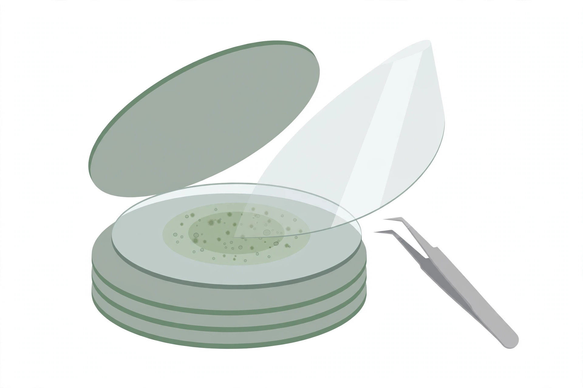
- Listeria species are enumerated on the 3M Petrifilm Environmental Listeria Plates (3M Petrifilm EL Plates), a sample-ready culture medium containing selective agents, nutrients, a cold-water-soluble gelling agent and a chromogenic indicator that facilitates Listeria colony detection
- The 3M Petrifilm EL Plate was designed to analyse environmental samples to help increase the efficiency of monitoring plant sanitation
- The presence of indicator Listeria such as Listeria innocua provides evidence that environmental conditions are suitable for the occurrence of Listeria monocytogenes
- The 3M Petrifilm EL Plate detects the majority of environmental Listeria: Listeria monocytogenes, Listeria innocua and Listeria welshimeri
- Samples are collected with a swab or a sponge, buffered peptone water is added as a repair broth, and a 3 mL sample is added directly to the plate and spread over a 42 cm² growth area
- The gelling agent is allowed to solidify, and the plates are incubated and then counted
- Intense red-violet colonies are indicative of Listeria; if colonies are present before the full 30 hour incubation but are not intense red-violet (for example grey or light pink), incubation continues up to 30 hours — colonies that do not turn intense red-violet by then are not interpreted as Listeria
- Studies performed by the University of Vermont, Silliker Laboratories and 3M have found the 3M Petrifilm EL Plate method to be comparable to traditional reference methods
Apparatus and materials
- 3M Petrifilm EL Plates 6447/6448 (3M Canada, Inc.) — store at or below 8 °C
- Plastic spreading device, Catalogue #6498
- Package insert, including instructions for use
- “Interpretation Guide” (available upon request)
- Stomacher, blender or equivalent
- Incubator, capable of maintaining 35 °C or 37 °C
- Pipettes or automatic pipettes having a nominal capacity of 1.0 to 5.0 mL
- Standard colony counter and/or illuminated magnifier
Reagents and diluents
- Appropriate sterile diluents: Letheen Broth or Neutralizing Broth if sanitizers are present; if not, the moistening agent can be sterile water or buffered peptone water (BPW)
- Repair broth: Buffered Peptone Water (the repair broth must be BPW)
- To prepare: dissolve 10.0 g of peptone, 5.0 g of sodium chloride (NaCl), 3.5 g of disodium phosphate and 1.5 g of potassium dihydrogen phosphate in 1.0 L of distilled water; final pH 7.2 ± 0.2. Dispense into appropriate glassware and autoclave at 121°C for 15 min.
Sample handling
- Sponge or swab samples are processed through the repair and plating steps immediately
- If a sample cannot be processed immediately, environmental samples may be stored in the refrigerator at 4–8 °C for up to 24 hours before repair and plating onto 3M Petrifilm EL Plates, without significant impact on numbers; quantification of Listeria is still possible
Preparation for analysis
- Have sterile diluent and repair broth ready, and disinfect the surface of the work area
- Place the 3M Petrifilm EL Plate on a flat surface and mark identifying sample information on the film
- Have the sterile sponge or swab ready, and label the collecting device with sample information
Procedure
Note: Each sample unit may be analysed individually, or the analytical units may be combined where the requirements of the applicable sampling plan can be met.
Preparation of sample (repair of injured Listeria)
Agitate liquids until the contents are homogeneous, to ensure a representative analytical unit
↓
Collect the environmental sample using a 3M Quick Swab or equivalent, a sponge, or another moistened collection device
↓
Aseptically add 2 mL (swab) or 5 mL (sponge) of sterile 20–30 °C buffered peptone water (repair broth) to the collected sample
↓
Vigorously mix, stomach or vortex the collected sample with the buffered peptone water for approximately one minute
↓
Allow the sample to remain at room temperature (20–30 °C) for 1 hour, up to a maximum of 1.5 hours, then vigorously mix again — this step is required for the repair of injured Listeria
Inoculation and incubation
Lift the top film and carefully inoculate 3.0 mL of sample onto the centre of the bottom film
↓
Roll the top film down onto the sample
↓
Distribute the sample evenly by gently placing the plastic spreader on the top film over the inoculum; do not press, twist or slide the spreader (the spreader is not necessary if the inoculum self-spreads)
↓
Leave the plate undisturbed for a minimum of 10 minutes to permit solidification of the gel
↓
Incubate the plates in a horizontal (upright) position with the clear side up, in stacks not exceeding 10 units, for 28 ± 2 h at 35 °C or 37 °C
Reading and interpretation
Count the plates promptly after the incubation period; if impossible to count at once, store the plates in the freezer (this is avoided as a routine practice)
↓
Count the red-violet colonies as Listeria, using a standard colony counter (a magnifier-illuminator may also be used to facilitate counting)
↓
If any colonies are present but are not intense red-violet (for example grey or light pink), continue incubating up to 30 hours
↓
At the maximum incubation time of 30 hours, colonies that do not turn intense red-violet are not counted as Listeria; the test is complete — do not exceed 30 hours, since incubation beyond the recommended time may yield ambiguous results
Note: Do not count colonies on the foam dam, since they are removed from the selective influence of the medium.
- There are three ways to read 3M Petrifilm EL Plates: quantitative, semi-quantitative and qualitative interpretation
- When colonies are present in large numbers, the plates may have many small, indistinct colonies and/or a pink/brown colour throughout — record the results as too numerous to count
- Estimates can be made on plates containing large numbers of Listeria by counting the number of colonies in one or more representative squares and determining the average number per square; multiply the average number by 42 to determine the total count per plate (the circular growth area is approximately 42 cm²)
- To isolate colonies for further identification, lift the top film and pick the colony from the gel
- Follow MFHPB-07 or MFHPB-30 to confirm colonies
Calculation
To determine the quantity of Listeria per sampled area, record:
- Area size sampled
- Volume of hydration fluid in the sampling device
- Volume of the buffered peptone water added
- Volume plated (3 mL per plate)
- Number of colonies counted
CFU / area = (Number of colonies × [volume of hydration fluid + volume of BPW] ÷ 3 mL) ÷ area sampled
Where,
– Number of colonies — number of colonies counted
– volume of hydration fluid — volume of hydration fluid in the sampling device
– volume of BPW — volume of the buffered peptone water added
– 3 mL — volume plated per plate
– area sampled — area size sampled
Precautions
- Do not use enrichment broth with the 3M Petrifilm EL Plate method
- It is the responsibility of each laboratory to ensure that the temperatures of the incubators or water baths are maintained at the recommended temperatures; where 35 °C is recommended in the text of the method, the incubator may be at 35 ± 1.0 °C
- Return unused plates to the foil pouch, seal the pouch by folding and taping the open end, and store the resealed foil pouch in a cool dry place
- Use plates within one month after opening the pouch; exposure of 3M Petrifilm Plates to temperatures above 25 °C and/or humidity above 50% RH can affect the performance of the plates
- Do not use plates that show orange or brown discoloration
- The expiration date and lot number are noted on each package of 3M Petrifilm Plates; the lot number is also noted on individual test films
- If the analyst uses any variations of the media listed here (either a commercially available product or one prepared from individual components), it is the responsibility of the analyst or Laboratory Supervisor to ensure equivalency
References
- Health Canada, Compendium of Analytical Methods, Volume 3 — Laboratory Procedure MFLP-11, February 2011: Enumeration of Listeria Species in Environmental Samples using 3M Petrifilm Environmental Listeria Plates
- McNamara, A.M., Kupski, B. and Zook, C. 2005. Evaluation of 3M Petrifilm Environmental Listeria Plate method for detection and enumeration of Listeria monocytogenes compared to the USDA-MPN and the VIDAS LIS methods. Technical poster presented at the 2005 University of Wisconsin-River Falls Microbiology symposium
- Groves, E. and Donnelly, C.W. 2005. Comparison of 3M Petrifilm Environmental Listeria Plate vs. Standard Methods in Detecting Listeria from Environmental Surfaces. Technical poster presented by University of Vermont at International Association for Food Protection (IAFP) annual meeting, August 2005, Baltimore, MD
- Groves, E., Nyachuba, D.G. and Donnelly, C.W. 2006. Comparison of 3M Petrifilm Environmental Listeria Plates with Selective Enrichment Methods for the Detection and Recovery of Listeria monocytogenes on Environmental Surfaces. Technical poster presented at the 2006 FDA Science Forum by E. Groves, D.G. Nyachuba and C.W. Donnelly, The University of Vermont, Department of Nutrition and Food Sciences
- Pagotto, F., Hébert, K. and Farber, J. 2011. Isolation of Listeria monocytogenes and other Listeria spp. from foods and environmental samples (MFHPB-30). In: Volume 2, The Compendium of Analytical Methods
- Warburton, D., Boville, A., Pagotto, F., Daley, E. and Chow, C. 2011. The Isolation of Listeria monocytogenes and other Listeria spp. from foods and environmental samples using palcam broth (MFHPB-07). In: Volume 2, The Compendium of Analytical Methods