SOP: Enumeration of Coliforms, Faecal Coliforms and E. coli in Foods Using the MPN Method
GIFSQ — Global Institute for Food Safety & Quality
1. Principle
Coliforms, faecal coliforms and Escherichia coli are used as indicator organisms: their presence in foods processed for safety can point to inadequate processing, post-processing contamination or microbial growth, and the presence of faecal coliforms and E. coli can point to faecal contamination. These terms are defined by the test itself — medium, incubation time and temperature — rather than by taxonomy. This method enumerates all three groups by the Most Probable Number (MPN) multiple-tube fermentation technique: three or more decimal dilutions of the sample are inoculated into broth tubes and incubated; gas production in Lauryl Sulfate Tryptose (LST) broth signals presumptive coliforms; gas in Brilliant Green Lactose Bile (BGLB) broth confirms coliforms; gas at 44.5 or 45 °C in EC broth confirms faecal coliforms; and typical nucleated, dark-centred colonies with or without metallic sheen on Levine’s Eosin Methylene Blue (L-EMB) agar, proven by biochemical tests, identify E. coli. The probable level of each group is estimated from the number of positive tubes using the standard MPN table (Appendix D of Volume 1). This method is not intended for isolating or enumerating pathogenic E. coli serotypes such as enterohaemorrhagic O157:H7, which do not give a positive faecal coliform reaction. This method replaces the June 2001 edition of MFHPB-19.
2. Apparatus required
- Covered water baths with circulating system (maintaining 44.5 °C and 45 °C; water level above the medium in immersed tubes)
- Calibrated, traceable thermometer
- Incubator at 35 °C
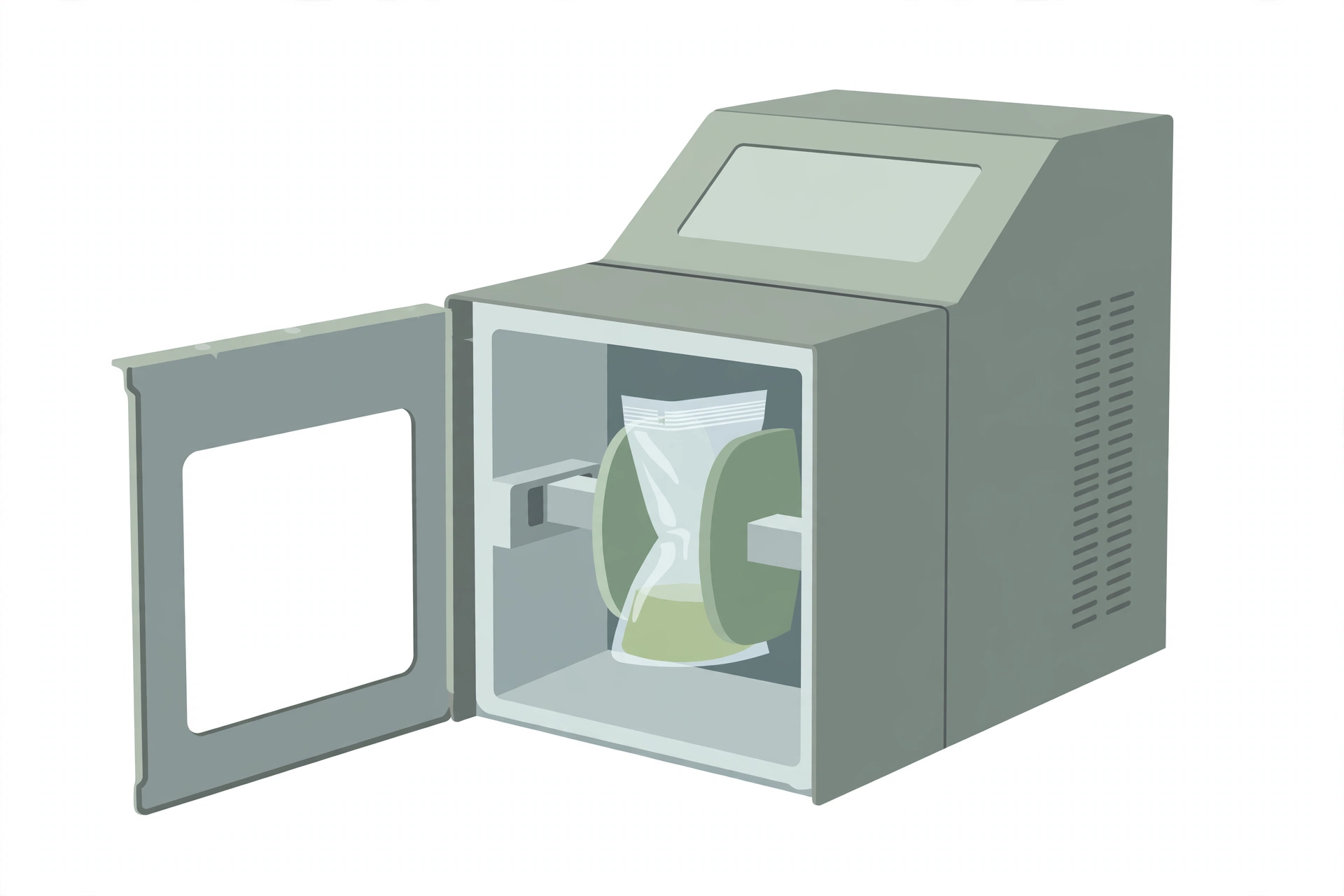
- Stomacher, blender or equivalent
- pH meter (distinguishing to 0.1 pH unit) or pH paper (distinguishing 0.3 to 0.5 pH units) covering pH 5.0 to 8.0
- Sterile pipettes (a separate sterile pipette for each transfer)
- Sterile disposable loops or sterile wood applicator sticks for tube-to-tube transfers
- Gas vials (Durham tubes) for broth tubes
- L-EMB or Endo agar plates
- Nutrient Agar (NA) or other non-selective agar
- UV light (366 nm) for examining EC-MUG tubes
3. Media and reagents
- 0.1% and 0.5% peptone water (0.5% peptone water for all shellfish dilutions)
- 0.1% peptone water — to prepare: dissolve 0.1 g of peptone in distilled water and make up to 100 mL.
- 0.5% peptone water — to prepare: dissolve 0.5 g of peptone in distilled water and make up to 100 mL (used for all shellfish dilutions).
- 2.0% aqueous sodium citrate (Na₃C₆H₅O₇·2H₂O), tempered to 40–45 °C — for cheese
- To prepare: dissolve 2.0 g of sodium citrate dihydrate (Na₃C₆H₅O₇·2H₂O) in distilled water and make up to 100 mL; temper to 40–45 °C before use.
- Lauryl Sulfate Tryptose (LST) broth, single and double strength
- Lauryl Sulfate Tryptose (LST) Broth: tryptose, tryptone or trypticase 20.0 g; lactose 5.0 g; dipotassium phosphate (K₂HPO₄) 2.75 g; monopotassium phosphate (KH₂PO₄) 2.75 g; sodium chloride (NaCl) 5.0 g; sodium lauryl sulphate 0.1 g per 1.0 L of distilled water; final pH 6.8 ± 0.2. Dissolve the ingredients in 1.0 L of distilled water (in 500 mL if double strength is required); dispense in 10.0 mL volumes into tubes containing gas vials; sterilize at 121°C for 15 min.
- Brilliant Green Lactose 2% Bile (BGLB) broth, dispensed in 10 mL volumes in tubes with gas vials
- Escherichia coli (EC) broth or EC broth with MUG (4-methylumbelliferyl-β-D-glucuronide), dispensed in 10 mL volumes in tubes with gas vials
- Levine’s Eosin Methylene Blue (L-EMB) agar or Endo agar
- Levine’s Eosin Methylene Blue (L-EMB) Agar: peptone 10.0 g; lactose 10.0 g; dipotassium phosphate (K₂HPO₄) 2.0 g; agar 15.0 g; eosin Y 0.4 g; methylene blue 0.065 g per 1.0 L of distilled water; final pH 7.1 ± 0.2. Add all the ingredients to distilled water; heat to boiling to obtain a complete solution; mix well; dispense as required (usually in 100–200 mL volumes); sterilize at 121°C for 15 min; avoid overheating, because the agar is likely to become soft and unsuitable for streaking.
- MacConkey agar
- MacConkey Agar: peptone 20.0 g; lactose 10.0 g; bile salts No. 3 1.5 g; sodium chloride (NaCl) 5.0 g; neutral red 0.03 g; crystal violet 0.001 g; agar 13.5 g per 1.0 L of distilled water; final pH 7.1 ± 0.2. Suspend the ingredients in 1.0 L of distilled water and heat to boiling with frequent swirling; autoclave at 121°C for 15 min and cool to 45–50°C; dispense the medium into plates and allow to dry with the lids partially removed.
- Nutrient Agar (NA) or other non-selective agar
- Nutrient Agar (NA): peptone 5.0 g; beef extract 3.0 g; agar 15.0 g per 1.0 L of distilled water; pH 6.8 ± 0.2. Suspend the ingredients in 1.0 L of distilled water and heat to boiling with frequent swirling; dispense into tubes or plates and autoclave at 121°C for 15 min; cool the tubes in a slanted position. Plates can be stored for up to 8 weeks at 4°C.
- 70% alcohol (shellfish shucking hand disinfection)
- To prepare: dilute 95% ethanol with distilled water to 70% (v/v) (for example, 74 mL of 95% ethanol made up to 100 mL).
- Iodophore solution or other suitable disinfectant (glove disinfection)
Commercially available media are prepared and sterilized according to the manufacturer’s instructions. If the analyst uses any variation of the listed media, the analyst or laboratory supervisor must ensure equivalency. Media formulae are given in Appendix G of Volume 1.
Supplies for confirmation (use option A or option B):
- Option A — IMViC media and reagents: tryptone (or tryptophane) broth with indole reagents; buffered glucose broth with Voges-Proskauer reagents and methyl red solution; Simmon’s Citrate (SC) agar
- Buffered Glucose Broth (MR-VP medium): peptone 5.0 g; glucose 5.0 g; dipotassium phosphate (K₂HPO₄) 5.0 g per 1.0 L of distilled water; final pH 7.5 ± 0.2. Add the ingredients to 1.0 L of distilled water; heat gently with stirring to obtain complete solution; dispense into test tubes; sterilize at 121°C for 15 min. The tryptone (or tryptophane) broth with indole reagents, Voges-Proskauer reagents, methyl red solution and Simmon’s Citrate agar are used as the method states.
- Option B — rapid identification kits or systems (such as API, Vitek or equivalent)
Control cultures (ATCC cultures or equivalent):
- Positive control(s): E. coli known to produce gas at 44.5 / 45 °C and to ferment lactose with typical reactions on L-EMB agar; if using EC-MUG, a strain known to produce β-glucuronidase
- EMB / IMViC negative control: Enterobacter aerogenes or an equivalent Gram-negative rod that does not give positive reactions on EMB and is indole-negative, methyl red-negative, Voges-Proskauer-positive and citrate-positive
- MPN broths negative control: Salmonella Berta or an equivalent Gram-negative rod that is gas-negative in the MPN broths and in the secondary EC broth
Some E. aerogenes strains produce a small gas bubble and false-positive reactions in the MPN broths (LST, BGLB and EC) — use S. Berta or an equivalent for those broths, and E. aerogenes or an equivalent for EMB agar and IMViC tests.
4. Sample preparation
- Keep sample units refrigerated (0–5 °C) or frozen before analysis, depending on the product (except shelf-stable foods); thaw frozen samples in a refrigerator or under time and temperature conditions that prevent microbial growth or death; analyse sample units as soon as possible after receipt — shellfish within 24 h of collection
- Each sample unit may be analysed individually, or analytical units may be combined where the sampling plan allows; combined analytical units should not exceed 500 g
- Have sterile peptone water ready; disinfect the working area; arrange LST broth tubes in rows of five and mark each with the sample unit and dilution
Shellfish (raw and processed):
- Use 0.5% peptone water for all dilutions; include only live animals for unfrozen shellfish; select 10 or more animals to obtain at least 200 g of meat and liquor
- Scrub the shellfish (including shell junctures) with a sterile stiff brush under running potable water — do not use faucets with aerators; drain in a clean container
- Disinfect hands or gloves (soap and water, potable-water rinse, then 70% alcohol rinse; or dip gloves in iodophore solution or suitable disinfectant and rinse with potable water; disposable gloves may be disinfected with 70% alcohol; a protective mail glove may be worn underneath), open each shellfish through the bill (not the hinge) with a sterile shucking knife, and collect meats and liquor in a sterile container
- Weigh at least 200 g of shellfish and liquor into a tared blender jar, add an equal amount of 0.5% peptone water, and blend 1–2 min — this homogenate is a 1 in 2 dilution
- For a 1 in 10 dilution, add 20 g of the homogenate to 80 g of peptone water and shake; shake each dilution 25 times through a 1-foot (30 cm) arc in about 7 s
- Prepare succeeding decimal dilutions with a separate sterile pipette per transfer; shake all dilutions immediately before transferring
- Within 2 min of blending, inoculate each of separate sets of five LST tubes per dilution: 10 mL of the 1 in 10 dilution into each of 5 tubes of double-strength LST; 1 mL of the 1 in 10 dilution into each of 5 tubes of single-strength LST; 1 mL of the 1 in 100 dilution into each of 5 tubes of single-strength LST (for fish products this scheme may be used optionally)
- The CSSP requires no more than 20 min from shucking to placing the inoculated tubes in the incubator
- Record results as MPN per 100 g of shellfish, using the Appendix D instructions
Water:
- Inoculate each of separate sets of five LST tubes per dilution: 10 mL of undiluted water into each of 5 tubes of double-strength LST (first row); 1 mL of undiluted water into each of 5 tubes of single-strength LST (second row); 0.1 mL of undiluted water into each of 5 tubes of single-strength LST (third row)
- Record results as MPN per 100 mL of water, using the Appendix D instructions
All other commodities:
- Agitate liquids or free-flowing materials until homogeneous; for solids, take the analytical unit from several locations within the sample unit
- Prepare a 1 in 10 dilution by aseptically blending 11 (10) g or mL of food into 99 (90) mL of the required diluent (see the table below); for a five-unit composite, blend 50 g or mL into 450 mL of diluent
- For fish products, an alternative is allowed: weigh 100 g, add 300 mL of 0.1% peptone water, blend 2 min (a 1 in 4 dilution); weigh 40 g of homogenate into 60 mL of 0.1% peptone water for the 1 in 10 dilution; inoculate as for shellfish above and express results as MPN/100 g
- Blend or stomach for the minimum time needed for a homogeneous suspension, not exceeding 2.5 min (avoid overheating); with foaming foods use low speed and remove the aliquot from below the liquid/foam interface
- Check the pH of the food suspension; if it is outside pH 5.5–7.5, adjust to 7.0 with sterile 1 N NaOH or 1 N HCl
- Let the 1 in 10 homogenate of dry foods stand at room temperature for 15 min; for all other foods continue without delay
- Prepare succeeding decimal dilutions with a separate sterile pipette per transfer; shake dilutions 25 times through a 1-foot (30 cm) arc in about 7 s, immediately before transferring
- Inoculate 1 mL of the 10⁻¹ dilution into each of five tubes of single-strength LST (first row); inoculate each succeeding row of single-strength LST with 1 mL of the next dilution
- For liquids: 1 mL of undiluted liquid into each of five tubes of single-strength LST (first row)
- Table option for solids: 10 mL of the 10⁻¹ dilution into 10 mL double-strength medium (represents 1 g)
Initial dilution by product type:
- Liquids (milk, water): pipette directly into LST and/or peptone water diluent — shake
- Viscous liquids: weigh into peptone water diluent — shake
- Water-soluble solids: weigh into peptone water diluent — shake
- Powders, meats: weigh into peptone water diluent — blend or stomach
- All cheese: weigh into previously warmed (40–45 °C) 2% aqueous sodium citrate — blend or stomach
- Spices: weigh into peptone water diluent — shake
- Shellfish, fish products: weigh into peptone water diluent — blend or stomach
The sample may be added to an empty stomacher bag, blender jar or dilution bottle with the diluent added before mixing.
5. Procedure
Set up growth controls and mix
- For each elevated-temperature water bath used, inoculate one LST broth tube with the MPN broths positive control and one with the MPN negative control; carry both into every medium used at each stage; set up an uninoculated tube of each medium at each step as a media control
- Mix inoculum and medium by gently shaking or rotating the tubes, avoiding air entrapment in the gas vials
↓
Incubate the LST tubes (presumptive coliform test)
- Incubate at 35 °C for 24 ± 2 h; examine for gas (a bubble or effervescence); record; for all gas-positive tubes begin the confirmed coliform, faecal coliform and E. coli tests as required
- Incubate gas-negative tubes (except raw shellfish and fish products) a further 24 ± 2 h; examine; add any new gas-positive tubes to the count and begin their confirmation tests
- No gas in any tube at the end of 48 ± 4 h (24 ± 2 h for raw shellfish and fish products) is a negative presumptive test
↓
Confirm coliforms in BGLB broth
- Transfer one loopful from each gas-positive LST tube (mix the tube first; avoid the pellicle) into a BGLB broth tube; sterile wood applicator sticks or other transfer devices may be used
- Mix gently without trapping air in the gas vials
- Incubate at 35 °C for 24 ± 2 h; examine for gas; record
- Incubate gas-negative tubes a further 24 ± 2 h; re-examine; add new gas-positive tubes to the count
- Gas within 48 ± 4 h is a positive confirmed coliform test
- Compute the MPN of confirmed coliforms per g (mL) of food (per 100 g of shellfish or fish products, or per 100 mL of water) from the number of gas-positive tubes using the Appendix D instructions; record
↓
Confirm faecal coliforms in EC broth
- Transfer one loopful from each gas-positive LST tube (mix first; avoid pellicles) into an EC broth tube — make this transfer at the same time as the BGLB transfer
- Mix gently without trapping air in the gas vials
- Incubate in a water bath at 45 °C for 24 ± 2 h (44.5 °C for shellfish and fish products); keep the bath water level at least 1 cm above the medium in the tubes
- Examine for gas; record; on the same day begin E. coli identification for all gas-positive tubes
- Incubate gas-negative tubes (except raw shellfish and fish products) a further 24 ± 2 h; examine; add new gas-positive tubes and begin their E. coli identification
- No gas at the end of 48 ± 4 h (24 ± 2 h for raw shellfish and fish products) is a negative presumptive test; gas within that time is a positive faecal coliform test
- If using EC-MUG broth, examine the tubes under UV light (366 nm): blue-green fluorescence is a positive presumptive E. coli test and those tubes may go on to E. coli confirmation
- Compute the faecal coliform MPN per g (mL) of food (per 100 g of shellfish and fish products, or per 100 mL of water) from the number of gas-positive tubes using the Appendix D instructions; record
↓
Identify E. coli
- Gently shake each gas-positive EC tube (or each fluorescing EC-MUG tube) and streak a loopful onto an L-EMB or Endo agar plate
- Incubate the plates at 35 °C for 18–24 h; look for typical non-mucoid, nucleated, dark-centred colonies with or without metallic sheen — indicative of E. coli
- The laboratory supervisor decides which dilutions and presumptive tube sets are confirmed, and how many colonies per plate are picked, to establish the final confirmed MPN count
- Well-isolated colonies: pick one typical colony and streak onto a non-selective agar such as NA (EMB or MacConkey may also be used); circle one other typical colony on the EMB plate before storing it at 4 °C in case the first colony does not confirm; incubate at 35 °C for 18–24 h and use these cultures for further confirmation
- Colonies not well isolated: pick two typical colonies and re-streak on EMB for discrete colonies; streak one well-isolated typical colony onto a non-selective agar such as NA (EMB or MacConkey may also be used); refrigerate the second EMB plate in case it is needed later; incubate and use these cultures for further confirmation
↓
Confirm by GIMViC or a rapid identification kit
GIMViC (or any commercially available testing system):
- Transfer inoculum from the non-selective plates into separate tubes of EC broth (G medium) and each IMViC medium — collectively the GIMViC media: G = secondary EC broth; I = tryptone broth; M and V = buffered glucose broth; C = Simmon’s Citrate agar; if GIMViC is not carried out within 96 h of inoculating the non-selective agar, prepare fresh plates or slants first
- Inoculate one tube of each GIMViC medium per isolate; inoculate IMViC positive and negative controls into each IMViC medium and MPN positive and negative controls into the secondary EC broth
- Gas at 44.5 °C or 45.0 °C (G): incubate the G medium (EC broth) in a water bath at 44.5 °C or 45.0 °C for 24 ± 2 h; examine for gas; if none, incubate a further 24 ± 2 h and re-examine; record
- Indole (I): incubate tryptone or tryptophane broth at 35 °C for 24 ± 2 h; add indole reagent per the manufacturer’s instructions — dark red in the alcohol layer is positive; orange probably indicates skatole (±); yellow is negative
- Methyl red and Voges-Proskauer (MVi): inoculate two tubes of buffered glucose broth; incubate at 35 °C for 48 ± 2 h; use the MR and VP reagents per the manufacturer’s instructions — VP is positive if an eosin pink colour develops after 5–10 min; MR is positive with a red colour and negative with a yellow colour
- Simmon’s Citrate (C): inoculate the SC agar slants with a straight needle using a light inoculum, avoiding carry-over of nutrients (carbon can give a false blue); incubate at 35 °C for 48 ± 2 h; visible growth with a colour change from green to deep blue is positive
- Characteristic GIMViC pattern for E. coli: Type I (typical) is gas-positive, indole-positive, methyl red-positive, Voges-Proskauer-negative, citrate-negative; Type II (anaerogenic) is gas-negative, indole-negative, methyl red-positive, Voges-Proskauer-negative, citrate-negative; commonly occurring coliforms can be differentiated using the coliform table below
- Differentiation of commonly occurring coliforms:
| Organism | Gas in EC broth at 44.5–45 °C | Indole test | Methyl red test | Voges-Proskauer test | Growth on citrate |
|---|---|---|---|---|---|
| Escherichia coli — Type I (typical) | + | + | + | − | − |
| Escherichia coli — Type II (anaerogenic) | − | − | + | − | − |
| Intermediates — Type I | − | − | + | −* | + |
| Intermediates — Type II | − | + | + | −* | + |
| Enterobacter aerogenes — Type I | − | − | − | + | + |
| Enterobacter aerogenes — Type II | − | + | − | + | + |
| Enterobacter cloacae — Irregular | − | − | − | + | + |
| Enterobacter cloacae — Type I | − | + | + | − | − |
| Enterobacter cloacae — Type II | + | − | + | − | − |
| Enterobacter cloacae — Type VI | + | − | − | + | + |
| Irregular — other types | Reactions variable |
Weak positive reactions are occasionally found.
– If the first isolate gives the characteristic pattern, the other isolate need not be tested; if it gives a non-characteristic IMViC pattern, test the second isolate’s GIMViC pattern; if both isolates fail to give the E. coli pattern, E. coli is absent from the primary EC broth tube they came from
Rapid identification kits:
- Use per the manufacturer’s instructions
↓
Compute the E. coli MPN per g (mL) of food (per 100 g of shellfish and fish products, or per 100 mL of water) from the number of tubes whose isolates give the characteristic GIMViC pattern or are confirmed as E. coli by a rapid kit, using the Appendix D instructions; record
6. Calculation
MPN values are obtained from the standard MPN tables and selection rules in Appendix D of Volume 1, using the number of gas-positive tubes (coliforms and faecal coliforms) or the number of tubes with E. coli-confirmed isolates:
- Foods (all other commodities): MPN per g (mL) of food
- Shellfish and fish products: MPN per 100 g
- Water: MPN per 100 mL
Where:
- MPN — the most probable number estimated from the positive-tube combination via the Appendix D tables
- g (mL) — per gram or millilitre of the food as analysed
- 100 g — per 100 g of shellfish or fish products
- 100 mL — per 100 mL of water
When assessing the presence of these indicator organisms in a sample, assess the results against the tolerance limits specified by government standards or guidelines, health agencies, or the laboratory’s in-house specifications. Established standards and guidelines are specifically linked to the method used to develop them
7. Precautions
- This method is not intended for isolating or enumerating pathogenic E. coli serotypes such as enterohaemorrhagic O157:H7 — many pathogenic serotypes do not give a positive faecal coliform reaction and would not be detected by this method
- Follow the manufacturer’s instructions and their safety precautions when using the UV light; examine negative EC-MUG control tubes under the UV light to make sure the tubes themselves do not fluoresce
- Maintain incubators and water baths at the stated temperatures: where 35 °C is given, the incubator may be at 35 ± 1.0 °C (except for shellfish, where it must be 35 ± 0.5 °C); 30 or 25 °C may be ± 1.0 °C; higher temperatures such as 43 or 44.5 °C must be held within 0.5 °C, and water baths within 0.2 °C, because higher temperatures can be lethal to the organisms being isolated
- Shake each dilution immediately before transferring to keep microorganisms uniformly distributed
- Use a separate sterile pipette for each transfer
- When transferring from positive LST tubes to BGLB or EC broth, avoid transferring the pellicle
- Do not trap air in the gas vials when mixing inoculum and medium
8. References
Health Canada, Compendium of Analytical Methods, Volume 2 — HPB Method MFHPB-19, April 2002: “Enumeration of coliforms, faecal coliforms and of E. coli in foods using the MPN method.”
- American Public Health Association. 1984. Recommended Procedures for the Examination of Sea Water and Shellfish; Fourth Edition. A.E. Greenberg and D.A. Hunt (eds.). American Public Health Association Inc., Washington, D.C.
- American Public Health Association. 2001. Compendium of Methods for the Microbiological Examination of Foods; Fourth Edition. Frances P. Downes and Keith Ito (eds.). American Public Health Association, Washington, D.C.
- American Public Health Association. 1992. Standard Methods for the Examination of Dairy Products; 16th Edition. R.T. Marshall (ed.). American Public Health Association Inc., Washington, D.C.
- American Public Health Association. 1998. Standard Methods for the Examination of Water and Waste Water; Twentieth Edition. Lenore S. Clesceri, A.E. Greenberg and A.D. Eaton (eds.). American Public Health Association, Inc., Washington, D.C.
- International Commission on Microbiological Specifications for Foods. 1978. Microorganisms in Foods; Their Significance and Method of Enumeration; Second Edition; University of Toronto Press.
- McGuire, O.E. 1964. Wood Applicators for the Confirmatory Test in Bacteriological Analysis of Water. Public Health Reports. 79: 812–814.
- Powers, E.M. and T.G. Latt. 1977. Simplified 48-Hour IMViC Test: an Agar Plate Method. Appl. Environ. Microbiol. 34: 274–279.