SOP: Determination of the Aerobic Colony Counts in Foods
GIFSQ — Global Institute for Food Safety & Quality
1. Principle
The Aerobic Colony Count (ACC) estimates the number of viable aerobic bacteria per g or mL of a food. A portion of the food is mixed with a plate count agar medium and incubated under defined conditions of time and temperature; each viable aerobic bacterium is assumed to multiply under those conditions and give rise to a visible colony that can be counted. The incubation temperature matches the growth-temperature requirements of the organism type being targeted. The method applies to psychrophilic, mesophilic and thermophilic aerobic bacteria in foods, for compliance assessment with Sections 4 and 7 of the Food and Drugs Act and other relevant federal regulations. It replaces MFHPB-18 of July 2015.
Definitions (as given in the method):
- Psychrophilic bacteria: an organism which grows optimally at or below 15°C, has an upper limit for growth at 20°C, and a lower limit of growth of 0°C or lower.
- Mesophilic bacteria: an organism whose optimum growth temperature lies within the range 20–45°C.
- Thermophilic bacteria: an organism whose optimum growth temperature is greater than 45°C.
2. Apparatus required
- Stomacher, blender or equivalent
- Incubator capable of maintaining the growth temperature required for the specific type of aerobic bacteria being enumerated
- Water bath capable of maintaining 45°C
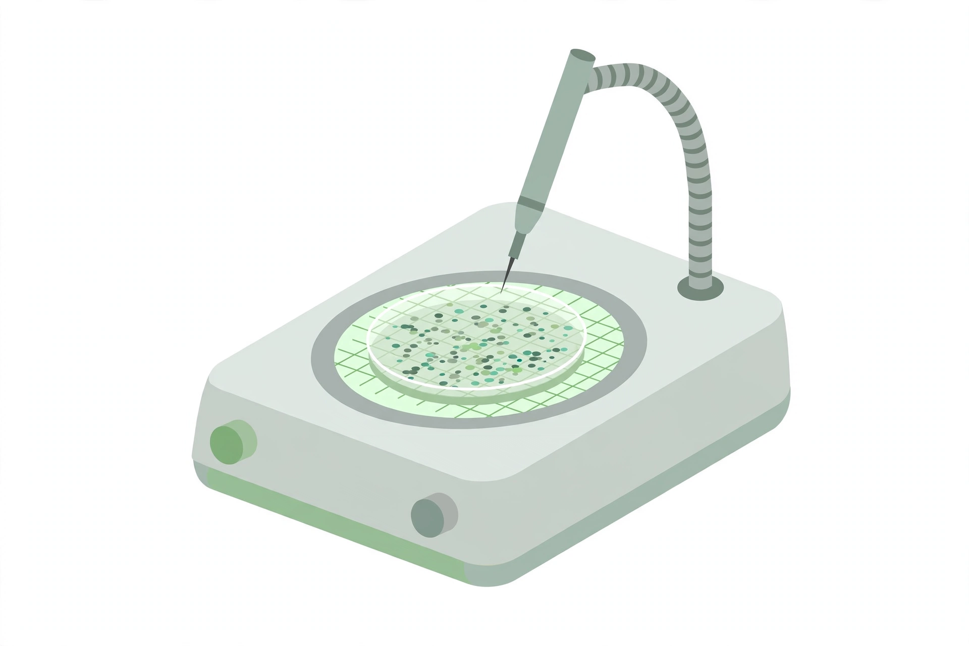
- Colony counting device (optional)
- Pipettes, sterile
- Petri dishes, sterile
3. Media and reagents
- Plate count agar (PCA)
- Plate Count Agar (PCA): tryptone 5.0 g; glucose 1.0 g; yeast extract 2.5 g; agar 15.0 g; distilled water 1.0 L; final pH 7.0 ± 0.2. Suspend the basal medium ingredients in 1.0 L of distilled water; heat to boiling to dissolve all ingredients; dispense into appropriate glassware and autoclave at 121 °C for 15 min. For shelf-stable milk products, addition of 1.0 mL of resterilised product to the tempered medium before pouring the plates may aid in the recovery of microorganisms.
- 0.1% peptone water diluent
- Peptone (0.1%) Water Diluent: peptone 1.0 g per 1.0 L of distilled water; final pH 7.0 ± 0.2. Suspend 1.0 g of peptone in 1.0 L of distilled water; mix thoroughly, and dispense into dilution bottles or tubes in amounts that will allow for volume lost due to autoclaving; autoclave at 121°C for 15 min.
- 2% sodium citrate, pre-warmed to 45°C (for cheese samples only)
- To prepare: dissolve 2 g of sodium citrate in distilled water and make up to 100 mL; pre-warm to 45°C before use.
- 0.1% 2,3,5-triphenyltetrazolium chloride (optional)
- To prepare: dissolve 0.1 g of 2,3,5-triphenyltetrazolium chloride in distilled water and make up to 100 mL.
- Suitable disinfectant for the working area
Commercially available media and reagents are prepared and sterilized according to the manufacturer’s instructions. See Appendix G of Volume 1 for the media formula.
4. Sample preparation
Initial dilution by food type (Table I of the method):
| Type of food | Preparation | Treatment |
|---|---|---|
| Liquids: milk, water, etc. | Pipette directly into Petri dishes and/or into peptone water diluent | Shake |
| Viscous lipids | Weigh into peptone water diluent | Shake |
| Solids: water-soluble solids, spices | Weigh into peptone water diluent | Shake |
| Solids: powder, meats, shellfish | Weigh into peptone water diluent | Blend or stomach |
| Cheese | Weigh into 2% sodium citrate pre-warmed at 45°C | Blend or stomach |
Dilution preparation
- Prepare sterile 0.1% peptone water diluent
- To obtain a representative analytical unit: agitate liquid or free-flowing materials until homogeneous; for a solid sample unit, take a portion from several locations within the unit
- Prepare a 1 in 10 (1:9) dilution of the food by aseptically blending or shaking 25 g or mL (the analytical unit) into 225 mL of the required diluent (see table above). With an analytical unit other than 25 g or mL, maintain the 1 in 10 dilution, e.g. 11 g in 99 mL, or 10 g in 90 mL
- If blending is used for homogenization, blending time must not exceed 2.5 min, to avoid overheating. With foods that tend to foam, blend at low speed and remove the aliquot from below the liquid/foam interface
- If shaking is used, shake the dilution bottles 25 times through a 30 cm arc in approximately 7 seconds
- Prepare succeeding decimal dilutions as required, using a separate sterile pipette for each transfer
- Shake all dilutions immediately before making transfers, to ensure uniform distribution of the microorganisms
5. Procedure
Determine which type of aerobic bacteria is being enumerated (psychrophilic, mesophilic and/or thermophilic). Analyze each sample unit individually.
↓
Handle the sample units: except for shelf-stable foods, keep sample units refrigerated or frozen in the laboratory before analysis, depending on the nature of the product; thaw frozen samples in a refrigerator, or under time and temperature conditions that prevent microbial growth or death; analyze sample units as soon as possible after receipt
↓
Prepare plate count agar, dispense in appropriate quantities, and sterilize
↓
Temper the prepared molten agar in a 45°C water bath, keeping the water level 1 cm above the level of the medium in the bottles
↓
Disinfect the surface of the working area with a suitable disinfectant
↓
Clearly mark the duplicate Petri plates for each sample and dilution
↓
Agitate each dilution bottle to resuspend material that may have settled during preparation
↓
Pipette 1 mL or 0.1 mL of the required dilutions into duplicate Petri plates. For products that tend to adhere to the bottom of the plates, add the inoculum to 1.0 mL of sterile diluent previously placed in the Petri plate
↓
Pour 12–15 mL of tempered agar into each plate and mix by rotating and tilting; allow to solidify. Pour the plates not more than 15 min after the dilutions were prepared
↓
Incubate the plates in the inverted position for 48 ± 4 h. Incubation temperature follows the growth-temperature requirements of the target organisms; suggested temperatures: psychrophilic bacteria 15–20°C, mesophilic bacteria 30–40°C, thermophilic bacteria 50–60°C. Plates for psychrophilic and thermophilic bacteria may be incubated up to 5 days. Other time/temperature combinations may be used if the laboratory has verified their suitability. Avoid crowding or excessive stacking of plates, to allow rapid equilibration with incubator temperature
↓
Count the colonies promptly after the incubation period
- Select plates with 25–250 colonies, including pinpoint colonies. If no counts fall within this range, plates nearest to the 25–250 range may be enumerated and the results reported as an estimate
- If plates contain spreading colonies, select a representative spreader-free portion of the plate and count the colonies in that area; estimate the total count of the whole plate by multiplying the area count by the reciprocal of the fraction counted — e.g. 30 colonies on 1/4 of the plate area gives 30 × 4 = 120 colonies for the whole plate
↓
Differentiate colonies from interfering food particles, if needed (optional):
- Option 1: for each dilution with interfering particles, prepare one extra plate and hold it under refrigeration as a control for comparison during counting (food particles such as meat or milk powder mimic colonies)
- Option 2: after incubation, flood the plates with 2 mL of 0.1% 2,3,5-triphenyltetrazolium chloride, rock gently from side to side to cover the whole area, pour off the excess, and keep the plates 3 h at room temperature in the inverted position. The bacteria reduce the indicator to a formazan that colours the colonies red, distinguishing them from food particles. Colonies cannot be picked for isolation after this treatment
↓
Record the results (see Calculation below)
6. Calculation
Concentration in the sample = total number of colonies on the plates selected for counting ÷ (total volume plated × dilution)
- Where,
- A, B = colony counts on the duplicate plates of one dilution
- C, D = colony counts on the duplicate plates of a second dilution (when used)
- DilutionAB = dilution of the first dilution pair (e.g. 10⁻¹)
- DilutionCD = dilution of the second dilution pair (e.g. 10⁻²)
Worked example 1 — one dilution in range. Two plates of a 10⁻¹ dilution show 34 and 42 colonies:
(34 + 42) CFU ÷ (2 mL × 10⁻¹ g/mL) = 380 CFU/g
Worked example 2 — two dilutions in range. Two plates of a 10⁻¹ dilution show 247 and 238 colonies, and two plates of a 10⁻² dilution show 26 and 25 colonies; the concentration is the weighted average from both dilutions:
[(247 + 238) + (26 + 25)] CFU ÷ [(2 mL × 10⁻¹ g/mL) + (2 mL × 10⁻² g/mL)] = 2436 CFU/g, rounded to 2400 CFU/g for reporting
Reporting rules
- Round the counts to two significant figures
- If the lowest dilution plated shows no colonies, record the count that would have been obtained from a single colony on one replicate plate, preceded by a “less than” (<) sign — e.g. two plates seeded with 1 mL of an undiluted liquid sample: < 0.5 CFU/g; with the lowest dilution 10⁻²: < 50 CFU/g
Reporting examples (Table II of the method):
| Observed average colony count | Dilution | Report as bacteria per g (mL) |
|---|---|---|
| 25–250, e.g. 144 | 1 in 1000 | 140,000 |
| Higher than 250, e.g. 440 | Highest dilution, 1 in 1000 | 440,000 E |
| Lower than 25, e.g. 15 | Lowest dilution, 1 in 1000 | 15,000 E |
| No count (0) | Lowest dilution, 1 in 1000 | Less than (<) 500 |
7. Precautions
- The laboratory supervisor must ensure the analysis is completed in accordance with the latest version of ISO/IEC 17025, “General Requirements for the Competence of Testing and Calibration Laboratories”
- Keep sample units refrigerated or frozen (except shelf-stable foods) and analyze them as soon as possible after receipt; thaw frozen samples only under conditions that prevent microbial growth or death
- Do not exceed 2.5 min of blending, to prevent overheating
- Pour the plates within 15 min of preparing the dilutions
- Do not crowd or excessively stack plates in the incubator
- Count plates with 25–250 colonies wherever possible; report out-of-range counts as estimates
- Do not pick colonies for isolation after treatment with 2,3,5-triphenyltetrazolium chloride
References
- American Public Health Association (APHA). 2015. Compendium of Methods for the Microbiological Examination of Foods. Fifth edition. Yvonne Salfinger and Mary Lou Tortorello (editors). Chapter 6. Culture Methods for Enumeration of Microorganisms. American Public Health Association Inc., Washington, D.C.
- Atlas, R.M. 1997. Handbook of Microbiological Media. Second edition. L.C. Parks (editor). CRC Press Inc.
- Solberg M. and B.E. Proctor, 1960. A technique utilizing 2,3,5-triphenyltetrazolium chloride for recognition of bacterial colonies in the presence of large numbers of food particles. Food Technology, 14 (7): 343–346.
- Holt J.G., Krieg N.R., Sneath P.H.A, Staley J.T. and S.T. Williams. 1994. Bergey’s Manual of Determinative Bacteriology. Ninth edition. W.R. Hensyl (editor). The Lippincott Williams and Wilkins Co., Baltimore.
Health Canada, Compendium of Analytical Methods, Volume 2 — HPB Method MFHPB-18, September 2020: “Determination of the Aerobic Colony Counts in Foods.”