SOP: Detection of Salmonella spp. in Foods Using the 3M Molecular Detection System Test Kit Version 2
GIFSQ — Global Institute for Food Safety & Quality
Principle
This method applies to the detection of Salmonella spp. to determine compliance with the requirements of Sections 4 and 7 of the Food and Drugs Act and/or other relevant federal regulations. Positive results must be confirmed with a cultural method. It has been validated for use in all foods except chocolate-based products, spices, powdered dairy products, and whole nuts.
The 3M Molecular Detection Assays use isothermal amplification of nucleic acid sequences combined with bioluminescence to detect the amplification of the target organism’s genetic material after 18-26 hours enrichment. Presumptive positive results are reported in real-time, while negative results are displayed after the assay is completed.
The chemistry is based on loop-mediated isothermal amplification (LAMP), a nucleic acid amplification technique. It allows the 3M Molecular Detection System to offer a simple sample preparation process with only two transfer steps. LAMP utilizes multiple primers that recognize distinct regions of the target Salmonella spp. gene and uses Bst polymerase, a unique enzyme with DNA strand-displacement activity, to enable continuous, rapid isothermal amplification.
The system uses bioluminescence technology to report the DNA amplification of the target organism in real-time. This involves a two-step enzymatic process in which pyrophosphate molecules, produced as a byproduct of the DNA amplification, are used to generate light. This light emission is then read by the 3M Molecular Detection Instrument and signals the detection of the target organism.
The unique method of bioluminescence detection, combined with the single-temperature amplification of LAMP used by the 3MMolecular Detection System, allows the target DNA to be amplified continuously, generating more than 10⁹ copies of target in as little as 15 min.
Apparatus
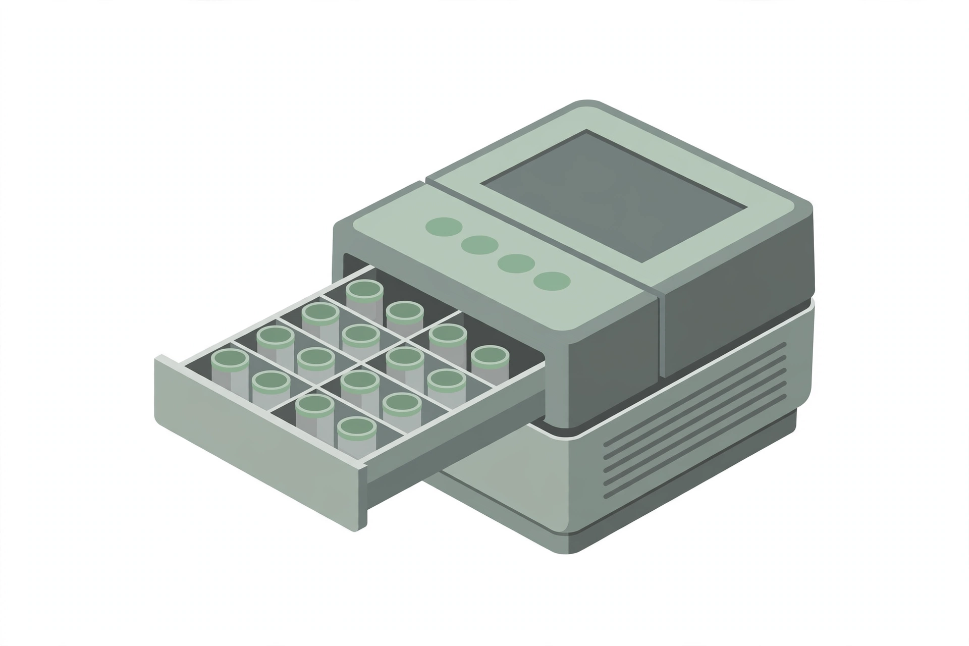
- MDS100 3M Molecular Detection Instrument and Accessory Kit (MDS100), which includes: the 3M Molecular Detection Speed Loader Tray, the 3M Molecular Detection Chill Block Insert, the 3M Molecular Detection Chill Block Tray, the 3M Molecular Detection Cap/Decap Tool—Lysis, the 3M Molecular Detection Cap/Decap Tool—Reagent, and empty lysis and reagent trays
- 3M Molecular Detection Assay 2 Salmonella spp. test kit (MDA2SAL96), which includes: 3M Lysis Solution (LS) tubes (96 – 12 strips of 8 tubes), 3M Salmonella spp. Reagent tubes (96 – 12 strips of 8 tubes), extra caps (96 – 12 strips of 8 caps), 3M Reagent Control (RC) (16 tubes), and the Quick Start Guide
- 3M Molecular Detection Heat Block Insert (MDSHBIN)
- 3M Molecular Detection Matrix Control (MDMC96) (optional)
- Incubator(s) that can be set at 37 ± 1°C and 41.5 ± 0.5°C
- Dry block heating unit that can fit the Insert (15 cm × 9.5 cm or 5.875″×3.75″) and reach temperatures of 100°C
- Single-channel 20 µL micropipette
- 8-Channel Multi-Channel 20 µL micropipette
- Sterile / barrier filter tips adaptable to 20 µL micropipettes
- Sterile stomacher bag
- Powder-free gloves
- Stomacher, blender, or equivalent
- Computer workstation
Note: It is the responsibility of each laboratory to ensure that the temperatures of the incubators or water baths are maintained at the recommended temperatures. The following applies to steps of the method which apply to growth only. Where a temperature of ≤ 37°C is recommended in the text of the method, the temperature may be ± 1. 0°C, e. g., 35 ± 1. 0°C. However, where higher temperatures are recommended, it is imperative that the incubators or water baths be maintained within 0. 5°C due to potential lethality of the higher temperatures on the microorganism(s) being isolated.
Reagents
The method formulates no media or reagents itself; the following are used as named and are prepared and sterilized according to the manufacturer’s instructions:
- Buffered Peptone Water (BPW)
- Basal medium: peptone — 10.0 g; sodium chloride (NaCl) — 5.0 g; disodium phosphate — 3.5 g; potassium dihydrogen phosphate — 1.5 g; distilled water — 1.0 L. Final pH 7.2 ± 0.2.
- To prepare: dissolve the ingredients in 1.0 L of distilled water ↓ dispense into appropriate glassware ↓ autoclave at 121 °C for 15 min.
- 3M Lysis Solution (LS) tubes
- 3M Salmonella spp. Reagent tubes
- 3M Reagent Control (RC) tubes
- 3M Matrix Control (MC) (optional)
- Sterile BPW (for the negative control)
Sample Preparation
Handling of sample units
- In the laboratory prior to analysis, except for shelf-stable foods, keep sample units refrigerated or frozen, depending on the nature of the product.
- Thaw frozen samples in a refrigerator, or under time and temperature conditions which prevent microbial growth or death.
- Analyze sample units as soon as possible after their receipt in the laboratory.
Preparation for analysis
- Have sterile BPW prepared and acclimatized to room temperature before use.
- Clean the surface of the working area with a suitable disinfectant.
- Turn on the block heater with the Heat Block Insert in, such that it can reach 100°C prior to analysis.
- Log in to the 3M Molecular Detection Software and turn on the 3M Molecular Detection Instrument.
Preparation of sample
- To ensure a representative analytical unit, agitate liquids or free-flowing materials until the contents are homogeneous; if the sample unit is a solid, obtain the analytical unit by taking a portion from several locations within the sample unit.
- Add 9 volumes (i.e., 225 mL) of room-temperature Buffered Peptone Water to each food sample (usually 25 g) in a stomacher bag.
- To reduce the workload, up to 15 analytical units may be composited into a single test sample (e.g., 375 g or mL) while maintaining the 1:10 dilution ratio.
- Blend, stomach, or vortex as required for thorough mixing.
- Incubate all samples for 18 – 26 h at 37 ± 1°C; incubate kefir for 18 – 26 h at 41.5 ± 0.5°C.
Procedure
DNA extraction — lysis step
Using one 3M Matrix Control (MC) per matrix type is suggested to determine if the matrix has the ability to impact the assay results.
↓
Allow the lysis solution (LS) tubes to warm up to room temperature by setting the rack on the laboratory bench for 2 h; invert the capped tubes to mix; one LS tube is required for each sample and a negative control sample
↓
Cut LS tube strips to the number needed; select the number of individual LS tubes or 8-tube strips needed; place the LS tubes in an empty rack
↓
Remove the enrichment broth from the incubator and gently agitate the contents
↓
To avoid cross-contamination, decap one LS tube strip at a time and use a new pipette tip for each transfer step
↓
Use the 3M Molecular Detection Cap/Decap Tool—Lysis to decap one LS tube strip — one strip at a time; set the tool with caps attached aside on a clean surface (caps can be retained in order to store lysis tubes if needed)
↓
Transfer 20 µL of sample into an LS tube; repeat until each individual sample has been added to a corresponding LS tube in the strip; repeat the decapping and transfer steps as needed for the number of samples to be tested
↓
When all samples have been transferred, transfer 20 µL of sterile BPW into an LS tube to act as the negative control (NC) (unless running own internal controls)
↓
Verify that the temperature of the 3M Molecular Detection Heat Block Insert is at 100 ± 1°C; place the uncovered rack of LS tubes in the Heat Block Insert and heat for 15 ± 1 min — the colour of the lysis solution should change from pink (cool) to yellow (hot)
↓
Remove the uncovered rack of LS tubes from the heating block and allow to cool in the 3M Molecular Detection Chill Block Insert for 5 ± 1 min — the lysis solution should change from yellow to pink, when cooled
↓
Remove the rack of LS tubes from the 3M Molecular Detection Chill Block Insert
Amplification and detection
Reagent tube strips can be cut to the desired number of tubes; select the number of individual Reagent tubes or 8-tube strips needed; one Reagent tube is required for each sample and the NC
↓
Place the Reagent tubes in an empty rack
↓
Select one Reagent Control (RC) tube and place it in the rack
↓
Transfer each sample lysate into individual Reagent tubes first, followed by the NC; hydrate the RC tube last; to avoid cross-contamination, decap one Reagent tube strip at a time and use a new pipette tip for each transfer step
Note: Care must be taken when pipetting LS, as carry-over of the protein residue from the bottom of the lysis tubes may interfere with amplification.
↓
Use the 3M Molecular Detection Cap/Decap Tool—Reagent to decap the Reagent tubes — one Reagent tube strip at a time; discard the cap
↓
Transfer 20 µL of sample lysate from the upper portion of the fluid in the LS tube into the corresponding Reagent tube; dispense at an angle to avoid disturbing the pellets; mix by gently pipetting up and down 5 times (using a multi-channel pipette can expedite this process)
↓
Repeat until all individual sample lysates have been added to a corresponding Reagent tube in the strip; repeat as needed for the number of samples to be tested
↓
Cover the Reagent tubes with the provided extra caps and use the rounded side of the 3M Molecular Detection Cap/Decap Tool—Reagent to apply pressure in a back-and-forth motion to ensure that the caps are tightly applied
↓
When all sample lysates have been transferred, transfer 20 µL of NC lysate into a Reagent tube to serve as a negative control; dispense at an angle to avoid disturbing the pellets; mix by gently pipetting up and down 5 times
↓
Transfer 20 µL of NC lysate into the RC tube to serve as a positive control; dispense at an angle to avoid disturbing the pellets; mix by gently pipetting up and down 5 times
↓
Load the capped tubes into a clean and decontaminated 3M Molecular Detection Speed Loader Tray; close and latch the Speed Loader Tray lid
↓
Review and confirm the configured run in the 3M Molecular Detection Software
↓
Click the Start button in the software and select the instrument for use — the selected instrument’s lid automatically opens
↓
Place the 3M Molecular Detection Speed Loader Tray into the 3M Molecular Detection Instrument and close the lid to start the assay — results are provided within 60 min, although positives may be detected sooner
Results and interpretation
An algorithm interprets the light output curve resulting from the detection of the nucleic acid amplification; results are analyzed automatically by the software and are color-coded
↓
A “Positive” or “Negative” result is determined by analysis of a number of unique curve parameters — negative samples will not give a zero reading, as the system and the 3M Molecular Assay Salmonella spp. amplification reagents have a background relative light unit (RLU)
↓
Presumptive positive results are reported in real-time, while “Negative” and “Inspect” results are displayed after the run is completed
↓
In the rare event of any unusual light output, the algorithm labels it as “Inspect” — repeat the assay for any “Inspect” samples beginning at the DNA extraction (lysis) step; if the result continues to be “Inspect”, proceed with cultural confirmation as described under Confirmation of Presumptive Positive Results
Confirmation of presumptive positive results
Commence with direct plating of the presumptive positive primary BPW enrichment to the selective agars as described in MFHPB-20, concurrently transferring an aliquot of the primary enrichment to the selective enrichments as described in MFHPB-20
↓
If the direct plating results confirm the presumptive positive result, the selective enrichment can be discontinued
↓
If the direct plating results are negative, continue with incubation of the enrichments, plating, isolation, and confirmation as described in MFHPB-20
Precautions
- The laboratory supervisor must ensure that completion of the analysis described in this method is done in accordance with the International Standards reference ISO/IEC 17025 (latest version): “General Requirements for the Competence of Testing and Calibration Laboratories” (as the method’s materials section states).
- It is the responsibility of the laboratory to ensure equivalency if any variations of the media formulations listed here are used (either product that is commercially available or made from scratch).
References
- Health Canada. 2020. MFLP-100: Detection of Salmonella spp. in Foods Using the 3M Molecular Detection System Test Kit Version 2. Volume 3. Compendium of Analytical Methods. Health Products and Food Branch.
- Notomi T, Okayama H, Masubuchi H, Yonekawa T, Watanabe K, Amino N, Hase T. 2000. Loop-mediated isothermal amplification of DNA. Nucleic Acids Res. 28(12): E63. 3.
- Francois P, Tangomo M, Hibbs J, Bonetti EJ, Boehme CC, Notomi T, Perkins MD, Schrenzel J. 2011. Robustness of a loop-mediated isothermal amplification reaction for diagnostic applications. FEMS Immunol. Med. Microbiol. 62(1):41-48.
- Kaneko H, Kawana T, Fukushima E, Suzutani T. 2007. Tolerance of loop-mediated isothermal amplification to a culture medium and biological substances. J Biochem Biophys Methods. 70(3):499-501.
- Kiddle G, HardingeP, Buttigieg N, Gandelman O, Pereira C, McElgunn CJ, Rizzoli M, Jackson R, Appleton N, Moore C, Tisi LC, Murray JAH. 2012. GMO detection using a bioluminescent real time reporter (BART) of loop mediated isothermal amplification (LAMP) suitable for field use. BMC Biotechnology. 12: 15.
- Plutzer J, Karanis P. 2009. Rapid identification of Giardia duodenalis by loop-mediated isothermal amplification (LAMP) from faecal and environmental samples and comparative findings by PCR and real-time PCR methods. Parasitol Res. 104(6):1527-1533.
- Nkouawa A, Sako Y, Li T, Xingwang C, Wandra T, Swastika K, Nakao M, Yanagida T, Nakaya K, Qiu D, Ito A. 2010. Evaluation of a loop-mediated isothermal amplification method using fecal specimens for differential detection of Taenia species from humans. J. Clin. Microbiol. 48(9):3350-3352.
- Gandelman OA, Church VL, Moore CA, Kiddle G, Carne CA, Parmar S, Jalal H, Tisi LC, Murray JAH. 2010. Novel bioluminescent quantitative detection of nucleic acid amplification in real-time. PloS ONE. 5(11):e14155.
- Reid A. 2009. Isolation of Salmonella from food and environmental samples (MFHPB-20), Volume 2. The Compendium of Analytical Methods.