SOP: Detection of Listeria spp. in Foods and Environmental Samples by the VIDAS Listeria Method
GIFSQ — Global Institute for Food Safety & Quality
This method (HPB Method MFHPB-29, March 2012) is applicable to the rapid detection of Listeria spp. for determining compliance with Sections 4 and 7 of the Food and Drugs Act. It has been validated for use in environmental samples and all foods except ready-to-eat meat and poultry, smoked fish, and yogurt, and replaces the December 2011 edition of this method. Note: although only certain food products are listed, the method may be suitable for other foods; other commodities must be validated as fit for purpose following the criteria in the Compendium of Analytical Methods, and the validation data forwarded to the Microbiological Methods Committee so the Application section can be expanded if the data fulfil MMC requirements.
1. Principle
The VIDAS LIS assay is an enzyme-linked fluorescent immunoassay (ELFA) run on the VIDAS system for the qualitative detection of Listeria spp. The internal surface of the Solid Phase Receptacle (SPR), a pipette tip-like disposable device, is pre-coated during production with anti-Listeria antibodies, and the assay configuration prevents non-specific reactions with the SPR. Reagents for an assay are held in a sealed multi-chambered strip.
All assay steps are performed automatically and sequentially by the VIDAS instrument. The sample is inoculated into the reagent strip and cycled in and out of the SPR for a specific length of time. Listeria antigens present in the sample bind to the anti-Listeria monoclonal antibodies coated on the interior of the SPR. Unbound sample material is washed away. Antibodies conjugated with alkaline phosphatase are then cycled in and out of the SPR and react with the Listeria antigen-antibody complexes already adsorbed to the SPR wall. Unbound conjugate is removed by washing.
The fluorescent substrate, 4-methyl-umbelliferyl-phosphate, is then cycled in and out of the SPR, where the enzyme conjugate converts the substrate to fluorescent 4-methyl-umbelliferone. The intensity of fluorescence is measured at 450 nm.
2. Apparatus required
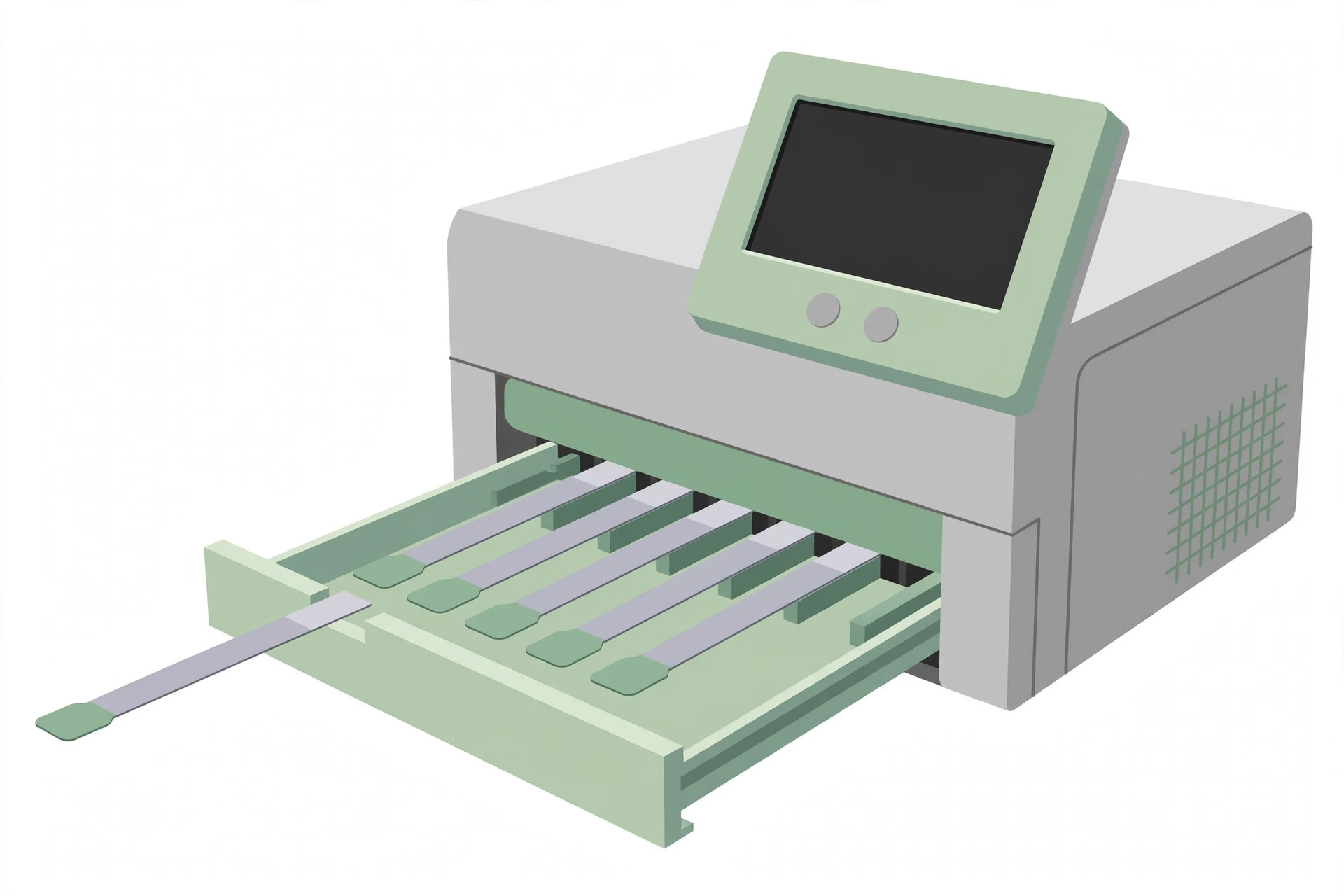
- VIDAS 30 (large capacity) or miniVIDAS (reduced capacity)
- Stomacher and stomacher bags, blender or equivalent
- Pipettor calibrated to dispense 500 µL and disposable tips
- Water bath at 95–100°C or equivalent system
- Incubators capable of maintaining 30°C and 35°C
3. Media and reagents
The following media are commercially available and are prepared and sterilized according to the manufacturer’s instructions (see Appendix G of Volume 1 and reference 7.1 for the formula of individual media):
- VIDAS Listeria Assay kit (Ref. 30 700): contains the necessary reagents, including standards and controls, for the analysis of 60 test samples. Available from bioMérieux Canada, Inc.
- Palcam broth
- Basal medium: peptone — 23.0 g; yeast extract — 5.0 g; lithium chloride (LiCl) — 10.0 g; esculin — 0.8 g; ammonium iron(III) citrate — 0.5 g; D(-) mannitol — 5.0 g; phenol red — 0.08 g; soybean lecithin — 1.0 g; Tween 80 — 2.0 g; distilled water — 1.0 L. Final pH 7.2 ± 0.2. Supplements (per 500 mL basal medium): polymyxin-B-sulphate — 5.0 mg; ceftazidime — 10.0 mg; acriflavine — 2.5 mg in 5.0 mL of distilled water (filter-sterilized).
- To prepare: suspend the basal medium ingredients in 1.0 L of distilled water and adjust the pH if necessary ↓ heat to boiling to dissolve completely ↓ autoclave at 121 °C for 15 min ↓ cool to 50 °C ↓ aseptically add the supplement solution to 500 mL of the cooled medium (double the amount for 1.0 L) ↓ mix thoroughly and dispense. Store under refrigeration in the dark, since acriflavine can photo-oxidize to form compounds inhibitory to Listeria spp.
- UVM 2
- Palcam agar (or equivalent agar)
- Basal medium: peptone — 23.0 g; starch — 1.0 g; sodium chloride (NaCl) — 5.0 g; yeast — 3.0 g; agar — 13.0 g; mannitol — 10.0 g; ferric ammonium citrate — 0.5 g; esculin — 0.8 g; dextrose — 0.5 g; lithium chloride (LiCl) — 15.0 g; phenol red — 0.08 g; distilled water — 1.0 L. Final pH 7.2 ± 0.2. Supplements (per 500 mL basal medium): polymyxin-B-sulphate — 5.0 mg; ceftazidime — 10.0 mg; acriflavine — 2.5 mg in 5.0 mL of distilled water (filter-sterilized).
- To prepare: suspend the basal medium ingredients in 1.0 L of distilled water and adjust the pH if necessary ↓ heat to boiling to dissolve completely ↓ autoclave at 121 °C for 15 min ↓ cool to 50 °C ↓ aseptically add the supplement solution to 500 mL of the cooled medium (double the amount for 1.0 L) ↓ mix thoroughly and dispense. Store under refrigeration in the dark, since acriflavine can photo-oxidize to form compounds inhibitory to Listeria spp.
- Oxford agar (or equivalent agar)
- Basal medium: Columbia blood agar base — 39.0 g; esculin — 1.0 g; ferric ammonium citrate — 0.5 g; lithium chloride (LiCl) — 15.0 g; agar — 2.0 g; distilled water — 1.0 L. pH 7.2 ± 0.2. Supplement per 10 mL (commercially available): cycloheximide — 400 mg; colistin sulphate — 20.0 mg; acriflavine — 5.0 mg; cefotetan — 2.0 mg; fosfomycin — 10.0 mg (dissolve in 10 mL of a 1:1 mixture of ethanol and distilled water; filter-sterilize).
- To prepare: suspend the basal medium ingredients in 1.0 L of distilled water ↓ heat to boiling with constant stirring to dissolve all ingredients ↓ autoclave at 121 °C for 15 min and cool to 45–50 °C ↓ aseptically add 10 mL of supplement and dispense ↓ store under refrigeration in the dark (acriflavine can photo-oxidize to form compounds inhibitory to Listeria spp.).
- Chromogenic media (optional): Rapid L. mono plates (Bio Rad Laboratories); Agar for Listeria according to Ottaviani & Agosti (ALOA)
4. Sample preparation
- Except for shelf-stable foods, keep sample units refrigerated or frozen in the laboratory before analysis, depending on the nature of the product. Thaw frozen samples in a refrigerator, or under time and temperature conditions which prevent microbial growth or death.
- Analyze sample units as soon as possible after their receipt in the laboratory.
- Have sterile Palcam broth ready, pre-warmed to approximately 35°C.
- Clean the surface of the working area with disinfectant.
- To ensure a representative analytical unit: agitate liquids or free-flowing materials until the contents are homogeneous. If the sample unit is a solid, obtain the analytical unit by taking a portion from several locations within the sample unit.
5. Procedure
Primary enrichment
Add 25 g or mL of the food (the analytical unit) to 225 mL of Palcam broth in a blender jar or stomacher bag. For composite samples, maintain a ratio of 1 part sample material to 9 parts of Palcam broth (see the Listeria policy for analytical unit sizes).
↓
For environmental samples: add the environmental sponge or large swabs to 100 mL of Palcam broth, or composite up to 10 sponges with 100 mL of Palcam broth per sponge (see MFLP-41). Place smaller environmental swabs (for example, cotton tip) in 10 mL portions of Palcam broth, or composite up to 10 swabs with 10 mL of Palcam broth per swab.
↓
Blend, stomach or vortex as required for thorough mixing and incubate at 35°C for 26–28 h.
Secondary enrichment
Agitate the Palcam broth sufficiently to mix and allow large food particles to settle. Transfer 1 mL of the Palcam broth to 9 mL of UVM 2. Incubate 26 (minimum) to 48 h at 30°C.
↓
Optional — weekend refrigeration of secondary enrichment cultures (UVM 2): UVM 2 cultures arising on Friday from samples analyzed on the preceding Wednesday or Thursday are refrigerated over the weekend. On the following Monday, the contents of the refrigerated UVM 2 cultures are resuspended and proceed as below.
↓
After incubation, transfer 2 mL of the enrichment broth into a tube and heat for 15 ± 2 min in a water bath at 95–100°C. Allow the tube to cool, then perform the VIDAS LIS test. Store the remaining enrichment broth at 2–8°C for later use in the confirmation of any VIDAS Listeria positive assay results.
Analysis on the VIDAS system
Remove the VIDAS LIS kit from the refrigerator. Remove the necessary components from the kit and allow them to come to room temperature (minimum 30 min). Return all unused components to storage at 2–8°C.
↓
In the space provided, label LIS Reagent Strips with the appropriate sample identification numbers.
↓
Follow the applicable instructions for entering master lot, sample, standard and control data, according to the operational instructions for the VIDAS system in use (VIDAS 30 or miniVIDAS). Components of different lots cannot be mixed and cannot be used past the expiry date.
↓
When a new kit lot is being used, first enter the specifications (the factory master calibration curve) into the system using the master lot entry card included with each kit.
↓
The standard must be calibrated a minimum of every 14 days and when a new kit lot is being used. If a standard needs to be tested, enter AS1@ for the sample identification. The standard must be situated at the beginning of the Work List, followed by the controls C1 and C2. The standard must be tested in duplicate, both with the same S1 designation, if it is to be stored in memory.
↓
Vortex standard, control and boiled samples. Pipette 500 ± 50 µL of standard, control or boiled sample into the centre of the sample well of a LIS Reagent Strip. All enrichment broths must be heated for 15 minutes at 95–100°C and cooled to room temperature prior to testing on VIDAS.
↓
Load the LIS Reagent Strips and LIS SPRs into the positions that correspond to the VIDAS section indicated by the Work List. Check that the colour labels with the three-letter assay code on the SPRs and the Reagent Strips match.
↓
Initiate the assay processing as directed in the VIDAS Operator’s Manual. The assay will be completed in approximately 45 minutes.
6. Interpretation of results
- Immunoassays producing relative fluorescence values (RFV) of ≥ 0.10 indicate the presumptive presence of Listeria spp. in the test sample.
- Confirmation: using the refrigerated secondary enrichment broth, presumptive positive results may be confirmed with the plating and confirmation steps of a cultural method (MFHPB-07 or MFHPB-30), as appropriate.
- If the results indicate that Listeria spp. have not been detected, it is understood that the presence of L. monocytogenes has also not been detected, since no species belonging to the Listeria genus were detected. However, if Listeria spp. are detected, regardless of the amount of colony screening or additional confirmation steps done, these results cannot be used to state that L. monocytogenes is absent. Report all species of Listeria that are identified.
7. Precautions
- It is the responsibility of each laboratory to ensure that the temperature of the incubators or water baths is maintained at the recommended temperatures. Where 35°C is recommended in the text of the method, the incubator may be 35 ± 1.0°C. Similarly, lower temperatures of 30 or 25°C may be ± 1.0°C. However, where higher temperatures are recommended, such as 43 or 45.5°C, it is imperative that the incubators or water baths be maintained within 0.5°C, due to potential lethality of higher temperatures on the microorganism being isolated.
- If the analyst uses any variations of the media listed here (either a commercially available product or one made from scratch), it is the responsibility of the analyst or Laboratory Supervisor to ensure equivalency.
- For further details, refer to the package insert of the VIDAS Listeria Assay kit.
References
Health Canada, Compendium of Analytical Methods, Volume 2 — HPB Method MFHPB-29, March 2012: “Detection of Listeria spp. in foods and environmental samples by the VIDAS Listeria method.”
Atlas, R.M. 1997. Handbook of Microbiological Media. Second edition. L.C. Parks (editor). CRC Press Inc.
Health Canada, Health Protection and Food Branch, Food Directorate. 2011. Policy on Listeria monocytogenes in Ready-to-Eat Foods.
Pagotto, F., Hébert, K., J. Farber. 2011. Isolation of Listeria monocytogenes and other Listeria spp. from foods and environmental samples (MFHPB-30). In: Volume 2. The Compendium of Analytical Methods.
Sewell, A.M., Warburton, D. W., Boville, A., Daley, E. F. and Mullen, K. 2003. A Comparison of the Health Products and Food Branch and the Enzyme Linked Fluorescent Assay Methods for the Isolation and Identification of Listeria spp. from Foods. Int. J. Food Micro. 81(2): 123-129.
Warburton, D., Boville, A., Pagotto, F., Daley, E. and Chow, C. 2011. The Isolation of Listeria monocytogenes and other Listeria spp. from foods and environmental samples using palcam broth (MFHPB-07). In: Volume 2. The Compendium of Analytical Methods.